. 2016 Oct 22;11(1):18.
doi: 10.1186/s13064-016-0073-y.

Contralateral migration of oculomotor neurons is regulated by Slit/Robo signaling

Affiliations

Contralateral migration of oculomotor neurons is regulated by Slit/Robo signaling

Brielle Bjorke et al. Neural Dev. .

Abstract

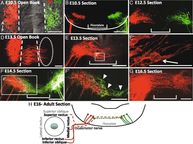
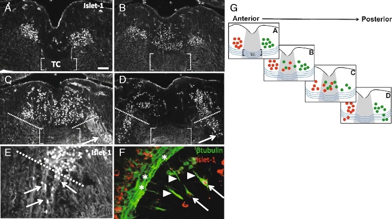
Background: Oculomotor neurons develop initially like typical motor neurons, projecting axons out of the ventral midbrain to their ipsilateral targets, the extraocular muscles. However, in all vertebrates, after the oculomotor nerve (nIII) has reached the extraocular muscle primordia, the cell bodies that innervate the superior rectus migrate to join the contralateral nucleus. This motor neuron migration represents a unique strategy to form a contralateral motor projection. Whether migration is guided by diffusible cues remains unknown.

Methods: We examined the role of Slit chemorepellent signals in contralateral oculomotor migration by analyzing mutant mouse embryos.

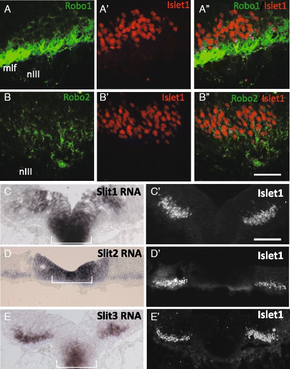
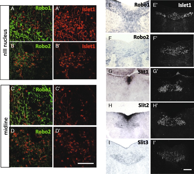
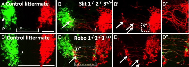
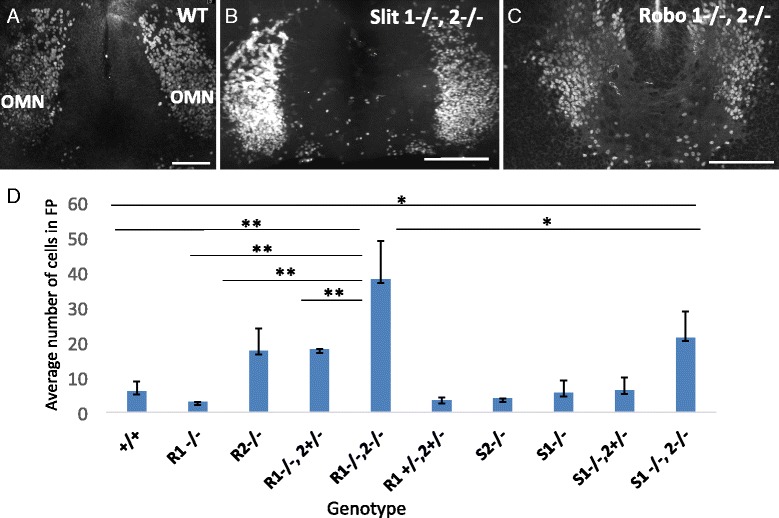
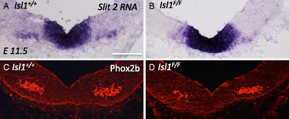
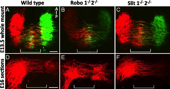
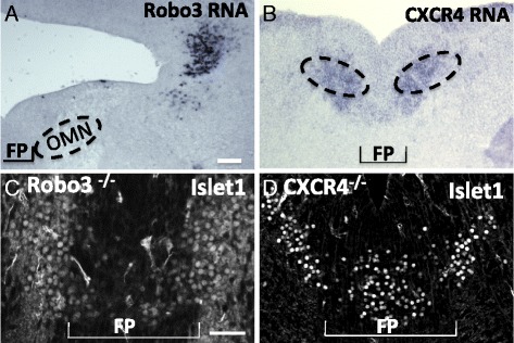
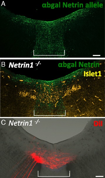
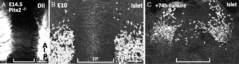
Results: We found that the ventral midbrain expresses high levels of both Slit1 and 2, and that oculomotor neurons express the repellent Slit receptors Robo1 and Robo2. Therefore, Slit signals are in a position to influence the migration of oculomotor neurons. In Slit 1/2 or Robo1/2 double mutant embryos, motor neuron cell bodies migrated into the ventral midbrain on E10.5, three days prior to normal migration. These early migrating neurons had leading projections into and across the floor plate. In contrast to the double mutants, embryos which were mutant for single Slit or Robo genes did not have premature migration or outgrowth on E10.5, demonstrating a cooperative requirement of Slit1 and 2, as well as Robo1 and 2. To test how Slit/Robo midline repulsion is modulated, we found that the normal migration did not require the receptors Robo3 and CXCR4, or the chemoattractant, Netrin 1. The signal to initiate contralateral migration is likely autonomous to the midbrain because oculomotor neurons migrate in embryos that lack either nerve outgrowth or extraocular muscles, or in cultured midbrains that lacked peripheral tissue.

Conclusion: Overall, our results demonstrate that a migratory subset of motor neurons respond to floor plate-derived Slit repulsion to properly control the timing of contralateral migration.

Keywords: Floor plate; Migration; Motor neuron; Oculomotor; Slit/Robo.


Figures

Fig. 1
Fig. 1
The oculomotor commissure is generated from E12.5 to E16.5 in the ventral midbrain. Oculomotor nuclei were back-labeled with peripheral application of DiI to the right oculomotor nerve (red) and DiO to the left oculomotor nerve (green) in mouse embryos on E10.5-16.5. The labeling is shown as either open book preparations revealing the anterior–posterior length of the oculomotor nucleus (A, D) or transverse sections of the midbrain (B, C, E, F, G), A, B. On E10.5, all oculomotor cell bodies were located on either side of the floor plate (A), ipsilateral to their nerve (B). C On E12.5, leading processes projected into the floor plate. D On E13.5, leading processes projected from the posterior half of the oculomotor nuclei across the midline toward the contralateral oculomotor nucleus. E Apparent cell bodies were located within the numerous leading processes within the floor plate (E’). F On E14.5, leading processes have crossed the floor plate to contact the contralateral nucleus. F’. In single focal planes by microscopy, contralateral cell bodies were located on the ventromedial aspect of the opposing nucleus as well as in the floor plate (arrow heads point to cell bodies outlined in green). G On E16.5 no cell bodies were located in the floor plate, and leading processes spanned the contralateral nucleus. H Schematic showing that the superior rectus extraocular muscle is innervated by contralateral oculomotor neurons and their midline axon fibers (dashed lines). Scale bars, 100 μm
Fig. 2
Fig. 2
On E13.5, Islet 1/2 positive neurons migrate across the midline independent of the existing commissure, with a small number of Islet positive neurons found in fibers projecting away from the nucleus. A-D. Transverse sections through the oculomotor nucleus on E13.5, shown anterior (A) to posterior (D), were antibody labeled for the motor neuron-specific transcription factor Islet1/2. The ventral tegmental commissure traveling through the floor plate is indicated (brackets in A-D). A In anterior sections, Islet1/2+ oculomotor neurons were located in distinct nuclei on either side of the floor plate, with no midline cell bodies visible (only non-specific blood vessel labeling is seen in the midline). B-D Large numbers of oculomotor neurons were located in the floor plate in a distinct stream above the commissure in intermediate (B) through posterior sections (C, D). In posterior sections, a subset of Islet1/2+ cells formed a stream toward the pial surface, moving outside of the bounds of the nucleus (marked with dashed white lines), apparently along the nerve fibers projecting to the ventral exit point (arrows in C, D). E Islet 1/2+ neurons were located in fibers projecting from the oculomotor nucleus toward the pial surface of the neural tube (arrows point to fibers projecting away from the oculomotor nucleus). F Sagittal section through the oculomotor nerve, projecting from ventral exit points (asterisks) toward the eye (arrow head points to peripheral oculomotor nerve fibers), shows Islet 1/2 positive cells located within the peripheral oculomotor nerve (arrows in F). G Schematic indicating the location of Islet1/2+ cell bodies in anterior to posterior sections. Right and left oculomotor nuclei are indicated by green and red colors respectively. The tegmental commissure is shown as blue curved lines traveling through the floor plate (gray color). Islet positive cells are located above the commissure. Abbrev: Tegmental commissure (TC). Scale bar100 μm
Fig. 3
Fig. 3
The guidance cues Slit1 and 2, and receptors Robo1 and 2, are in position to prevent oculomotor migration across the floor plate on E10.5. Coronal sections were taken through the posterior midbrain on E10.5 to determine Slit and Robo expression. Following protein (Robos) or mRNA (Slits) labeling, the same or adjacent section was labeled for Islet1/2 to co-localize expression to oculomotor neurons (A’-B’, A”, B”). A, B Robo1 and 2 protein was found in the ventral midbrain and in the oculomotor nerve fibers (nIII). Expression was co-localized with Islet1/2 indicating Robo1 and Robo2 expression in oculomotor neurons (A”,B”). Robo1 antibody also strongly labels the adjacent medial longitudinal fasciculus (mlf). C-E In situ hybridization for Slit1, 2, and 3 mRNA. Slit 1, 2 and 3 expression was localized to the floor plate. Expression of Slit2 and 3 co-localized to Islet 1/2+ neurons (D’, E’). Scale bars: B”, 50 μm, applies to Robo antibody labels; C’, 100 μm, applies to Slit in situs
Fig. 4
Fig. 4
Slit and Robo remain in position to regulate floor plate crossing on E14.5. Coronal sections through the caudal oculomotor nucleus on E14.5 were labeled with Robo1 or 2 antibodies, or hybridized to Robo or Slit mRNA probes, followed by antibody labeling for Islet1/2 of the same section (for Robo antibodies), or adjacent sections (for in situ hybridization). A, B Within the nucleus, Robo1 and Robo2 was localized to Islet1/2+ cells indicating Robo expression by oculomotor neurons. C-D. In the midline of the caudal midbrain, Robo1 antibody labeling could be seen on some migrating neurons, while Robo2 labeling was less intense and variable. E, F In situ hybridization for Robo1 and 2 mRNAs showed labeling that overlapped with the nuclei, and also bridged across the midline in the area of migrating neurons. G-I. Slit mRNA expression by in situ hybridization, compared to Islet antibody labeling in adjacent sections. Slit1 and 2 continued to be expressed by floor plate cells on E14.5, while there was very little Slit3. Slit2 and Slit3 transcript was localized to the oculomotor nuclei as well as overlapping with the motor neurons migrating across the midline (H, I). Scale bars: D’, 50 μm, applies to Robo antibody labels; I’, 100 μm, applies to in situs
Fig. 5
Fig. 5
Motor neuron cell bodies migrate prematurely into and across the floor plate in Slit and Robo mutants. A-D. On E10.5, DiI and DiO were applied to the left and right (respectively) peripheral oculomotor nerves to back-label the oculomotor nucleus, as well as the leading process and somata directly connected to the peripheral nerve. Open book preparation of the mouse midbrain, with anterior up. A, C On E10.5 in wild type littermate controls, oculomotor somata and axons remained ipsilateral to the nucleus. Leading projections are rarely seen projecting from the oculomotor nuclei (asterisk in A, C). B, D Slit1 -/-,-2-/- mutants or Robo1 -/-,2-/- mutants had numerous leading processes projecting into and across the floor plate. Cell bodies were found in the ventral region of the contralateral nucleus (yellow, arrows in B, D). Bulges in leading processes appeared to be cell bodies migrating across the floor plate (arrows in B’,D’). Leading processes looped into and across the floor plate in Slit mutants (B”). Robo mutants displayed more fasciculation by leading processes traversing through the floor plate (yellow color in box (D”). (wild type, n = 6; Slit1 -/-, 2-/-, n = 8; Robo1 -/-,2-/-, n = 9) Scale bars, 100 μm
Fig. 6
Fig. 6
Cells migrating through the posterior midbrain in Slit and Robo mutants are motor neurons. A-C To identify and quantify migrating neurons that have separated from the oculomotor nucleus in Slit and Robo mutant mice on E10.5, open book preparations were antibody labeled with Islet1/2. In wildtype controls, few Islet1/2+ cells were found within the floor plate (A), while numerous Islet1/2+ cells were seen in the floor plate between the left and right oculomotor nuclei in both Slit and Robo mutants (B, C). D The average number of Islet 1/2+ cells located medial to the oculomotor nuclei were counted for each genotype (2 or more litters per genotype). The number of cells medial to the oculomotor nucleus in Robo1-/-,2-/- and Slit1-/-, 2-/- mutants is significantly more than wildtype controls. There are more Islet 1/2+ cells in the floor plate in Robo mutants compared to Slit mutants (D). Abbrev. Anterior (A), posterior (P). Scale bars, 100 μm, Error bars indicate standard deviation, **P < 0.01, *P < 0.05. (control, n = 5; Robo1 -/-, n = 2; Robo2 -/-, n = 4; Robo1 -/-,2+/-, n = 2; Robo1-/-,2-/-, n = 6; Robo1+/-, 2+/-, n = 5; Slit1-/, n = 2; Slit2-/-, n = 3; Slit1-/-,2+/-, n = 4; Slit1-/-,2-/-, n = 9)
Fig. 7
Fig. 7
Loss of slit 2 in motor neurons is not sufficient to cause premature migration. A, B To confirm a loss of Slit2 expression in the oculomotor nucleus in Islet1 mutant embryos, in situ hybridization was performed in the ventral midbrain on E11.5. C, D. Location of the oculomotor nucleus was determined by Phox2b expression (an Islet-independent transcription factor expressed in motor neurons). A In control embryos, Slit2 RNA is found in the floor plate and co-localized to Phox2b positive cells (C). B In the Islet1 mutant midbrain, Slit2 expression is retained in the floor plate, but is lost from the oculomotor nucleus (D). Phox2b positive neurons were clustered on either side of the floor plate, but not within the floor plate in both control and Islet1F/F mutants (B) indicating oculomotor cell bodies have not migrated into the floor plate (n = 4). Scale bar, 100 μm
Fig. 8
Fig. 8
Slit and Robo mutants generate a normal oculomotor commissure. A-C Open book preparation of DiI and DiO back-labeled oculomotor nuclei. On E13.5, leading processes reached the contralateral nucleus. Neuron cell bodies, seen as bulges in the leading process, were in the midline (A, wild type control, Slit littermate). Loss of Slits or Robos (B, C) did not appear to reduce the number of leading processes or cells migrating through the floor plate. D-F. DiI back-label of the oculomotor nucleus on E16. 200 um coronal sections along the plane of the oculomotor nerve were compared by z-stacked confocal images. The oculomotor commissure was similar in thickness to wild type (D) in Slit and Robo mutants (E, F). (n = 3; control, Robo and Slit mutants) Scale bars, 100 μm
Fig. 9
Fig. 9
Robo3 or CXCR4 function is not required for oculomotor migration into the floor plate. A, B. To determine if Robo3 or CXCR4 are in position to repress Slit/Robo signaling in situ hybridization of RNA was performed on E14.5 midbrain sections. Robo3 was not expressed in the ventral midbrain on E14.5 (A). However, CXCR4 was expressed in the ventral midbrain in both the floor plate and regions lateral to the floor plate (B). C, D To identify migration patterns of oculomotor neurons on E13.5 in Robo3 and CXCR4 mutants, sections of the posterior midbrain were antibody labeled with Islet1/2. Islet1/2+ cells migrated into the floor plate in both Robo3 (C) and CXCR4 mutants (D) indicating neither Robo3 nor CXCR4 are required for migration oculomotor migration across the floor plate. (Robo3, n = 2; CXCR4, n = 4) Scale bar, 100 μm
Fig. 10
Fig. 10
Netrin1 is not required for oculomotor migration into the floor plate. A, B To locate Netrin expression during oculomotor migration, the mutant allele Netrin1lacZ was labeled with anti beta-gal in Netrin1-/- (i.e. lacZ/lacZ) mutant mice. On E13.5, Netrin1 was expressed by the floor plate and ventricular layer of cells in ventral midbrain (A), adjacent to Islet1/2+ motor neurons located in the nucleus and in the floor plate (B). C. DiI back-label from nIII in a Netrin1-/- embryo on E13.5. A 200 um coronal section along the plane of the oculomotor nerve was imaged by z-stacked confocal images. Leading processes projected from the oculomotor nucleus toward the contralateral nucleus (n = 3). Scale bar, 100 μm
Fig. 11
Fig. 11
Oculomotor midline migration is independent of peripheral signals. A. Open book preparation of the midbrain in a mouse lacking extraocular muscles in Pitx2 mutant mice, with the oculomotor nucleus back labeled with DiI. On E14.5, a commissure originating from the posterior half of the oculomotor nucleus crossed the floor plate (n = 3). B, C. Explant culture of isolated midbrain tissue. E10.5 midbrains were dissected to remove all peripheral tissue, including nIII, then cultured in an open book preparation in collagen gel for 72 h. Cultured tissue was then labeled with Islet1/2 antibody. Anterior is up; floor plate indicated by bracket. B. Dissected tissue at the onset of culture period showed Islet1/2+ cell bodies on either side of the floor plate. C. Following incubation for 72 h, a posterior subset of oculomotor neurons migrated into the midline (n = 4). Brackets indicate floor plate region. Scale bars, 100 μm

References

    1. Granet DB, Khayali S. Amblyopia and strabismus. Pediatr Ann. 2011;40:89–94. doi: 10.3928/00904481-20110117-08. - DOI - PubMed
    1. Puelles L, Privat A. Do oculomotor neuroblasts migrate across the midline in the retal rat brain? Anat Embryol (Berl) 1977;150:187–206. doi: 10.1007/BF00316650. - DOI - PubMed
    1. Puelles L. A Golgi-study of oculomotor neuroblasts migrating across the midline in chick embryos. Anat Embryol (Berl) 1978;152:205–215. doi: 10.1007/BF00315925. - DOI - PubMed
    1. Hogg ID. Observations of the development of the nucleus of Edinger-Westphal in man and the albino rat. J Comp Neurol. 1966;126:567–584. - PubMed
    1. Puelles-Lopez L, Malagon-Cobos F, Genis-Galvez JM. The Migration of Oculomotor Neuroblasts Across the Midline in Chick Embryo. Exp Neurol. 1975;47:459–469. doi: 10.1016/0014-4886(75)90078-3. - DOI - PubMed

MeSH terms

LinkOut - more resources