. 2016 Dec:57:258-269.
doi: 10.1016/j.neuro.2016.10.010. Epub 2016 Oct 19.

Pb exposure prolongs the time period for postnatal transient uptake of 5-HT by murine LSO neurons

Affiliations

Pb exposure prolongs the time period for postnatal transient uptake of 5-HT by murine LSO neurons

Sunyoung Park et al. Neurotoxicology. 2016 Dec.

Abstract

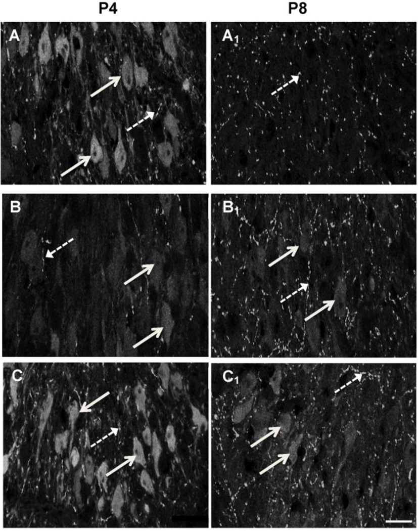
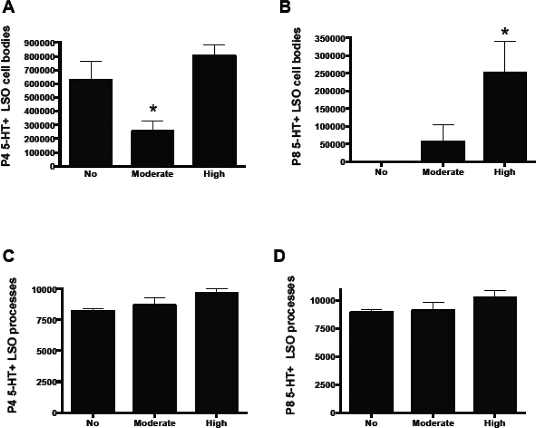
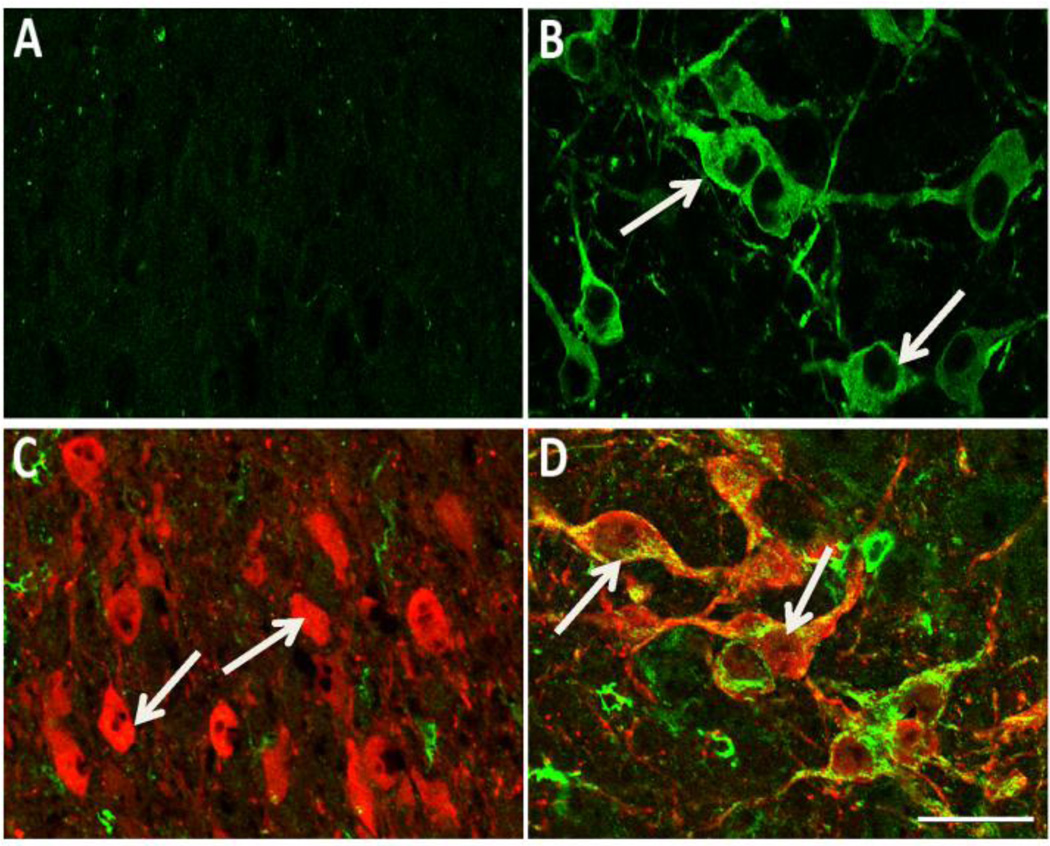
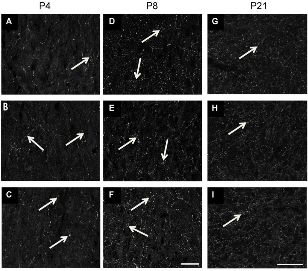
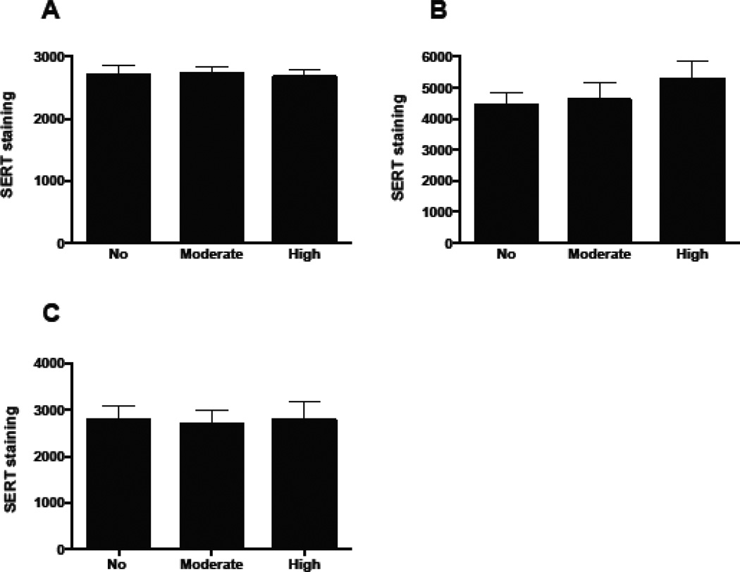
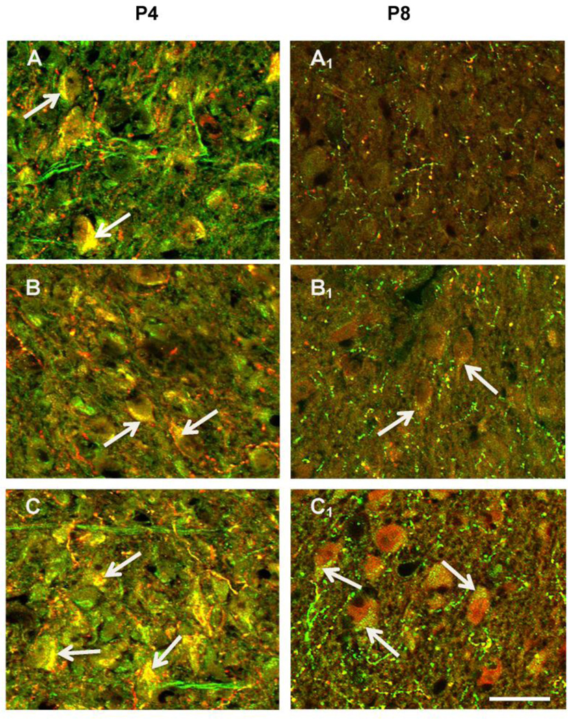
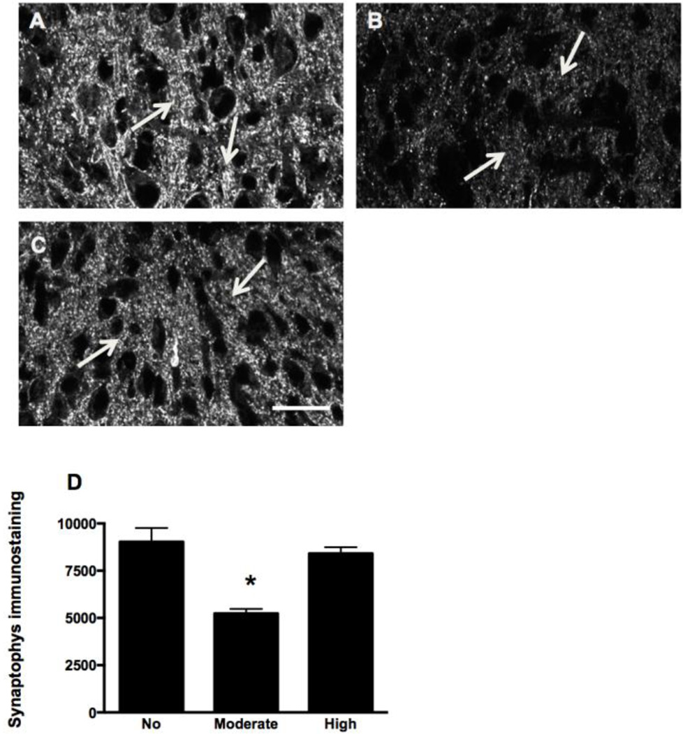
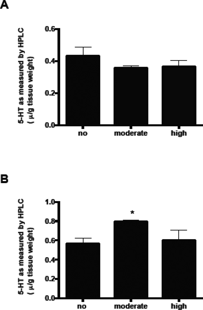
Pb exposure is associated with cognitive deficits including Attention Deficit Hyperactivity Disorder (ADHD) in children and alters auditory temporal processing in humans and animals. Serotonin has been implicated in auditory temporal processing and previous studies from our laboratory have demonstrated that developmental Pb decreases expression of serotonin (5-HT) in the adult murine lateral superior olive (LSO). During development, certain non-serotonergic sensory neurons, including auditory LSO neurons, transiently take up 5-HT through the serotonin reuptake transporter (SERT). The uptake of 5-HT is important for development of sensory systems. This study examines the effect of Pb on the serotonergic system in the LSO of the early postnatal mouse. Mice were exposed to moderate Pb (0.01mM) or high Pb (0.1mM) throughout gestation and postnatal day 4 (P4) and P8. We found that Pb exposure prolongs the normal developmental expression of 5-HT by LSO neurons and this is correlated with expression of SERT on LSO cell bodies. The prolonged expression of 5-HT by postnatal LSO neurons is correlated with decreased synaptic immunolabeling within the LSO. This Pb-associated decrease in synaptic density within the LSO could contribute to the auditory temporal processing deficits and cognitive deficits associated with developmental Pb exposure.

Keywords: Auditory system; Development; Lead acetate; Serotonin; Serotonin reuptake transporter (SERT); Superior olivary nuclei.


Figures

Figure 1
Figure 1
5-HT immunofluorescence labeling in somata and processes within the LSO of postnatal day (P) 4 and P8 mice. A–C (P4): A) P4 LSO neurons are immunopositive for 5-HT (solid arrows) and some LSO processes are also immunopositive (broken arrows). B) P4 LSO neurons are immunopositive for 5-HT following moderate Pb treatment (arrows), but the immunoreactivity is decreased compared to the no Pb controls. 5-HT+ LSO processes are similar to the no Pb control (broken arrows). C) P4 high Pb LSO neurons have similar 5-HT immunoreactivity compared to the no Pb controls (arrows). Broken arrows=5-HT+ LSO processes. A1–C1 (P8): A1) By P8, control LSO neurons are no longer immunopositive for 5-HT (arrows) although there are many immunopositive LSO processes (broken arrows). Both the moderate (B1) and high (C1) Pb exposure results in 5-HT+ LSO neurons (arrows) and processes (broken arrows). Thus Pb extends the developmental window in which LSO neurons are immunopositive for 5-HT through P8. Bar=15 µ.
Figure 2
Figure 2
Quantification of 5-HT immunostaining in LSO cell bodies and processes. At P4, moderate Pb exposure significantly decreases 5-HT expression in LSO neurons compared to controls while high Pb results in 5-HT expression that is similar to controls (A). At P8, the high dose of Pb results in significantly more 5-HT immunoreactivity in LSO cell bodies compared to controls, while the moderate dose of Pb shows a trend towards increased immunostaining (B). Neither the moderate nor the high dose of Pb affected the 5-HT immunostaining of LSO processes at either P4 (C) or P8 (D). Graphs represent mean IOD ± SEM. *p<0.05; ANOVA with Dunnett’s post hoc test.
Figure 3
Figure 3
LSO neurons of P4 mice do not express tryptophan hydroxylase (TPH) or monoamine oxidase-A (MAOA). A, B: The LSO (A) does not contain any TPH (green) immunopositive somata unlike neurons in the raphe pallidus (B) where there are many immunopositive neurons (arrows). C, D: Double immunolabeling of 5-HT (red) and MAOA (green) shows that the 5-HT positive neurons in the LSO (C) do not contain any MAOA positive somata (arrows), unlike neurons in the raphe pallidus where there is obvious double label (yellow) within many neurons (D; arrows). Scale bar = 30 µ.
Figure 4
Figure 4
SERT immunolabeling in LSO. Pb does not result in significant changes in total SERT expression within the LSO at either P4 (A–C) or P8 (D–F) or P21 (G–I) (arrows). Controls (A, D, G), Moderate Pb (B, E, H), High Pb (C, F, I). Scale bar = 30µ (A–F), Scale bar = 50µ (GI).
Figure 5
Figure 5
Quantification of SERT immunostaining confirms that Pb does not significantly affect total SERT immunoreactivity in the LSO. However, the amount of SERT immunoreactivity is higher at P8 (B) compared to P4 (A) and P21 (C). Graphs represent mean IOD ± SEM. *p<0.05; ANOVA with Dunnett’s post hoc test.
Figure 6
Figure 6
Micrographs of LSO double-labeled for 5-HT (red) and SERT (green) in the LSO in P4 and P8 mice. Neurons in LSO at P4 are double labeled for both 5-HT and SERT (arrows) in control, moderate Pb, and high Pb animals. By P8, control neurons are no longer double-labeled (A1), but LSO neuron in both the moderate (B1) and high Pb (C1) treatment groups continue to be double-labeled for both 5-HT and SERT. Scale bar = 15µ.
Figure 7
Figure 7
Pb exposure decreases synaptophysin (SYP) immunolabeling in the LSO of P8 mice. A–C: Immunoreactivity for SYP (arrows) decreases with moderate Pb (B) compared to control (A). High Pb (C) does not result in changes in the SYP expression in the P8 LSO. D: Quantification of SYP immunostaining in the LSO confirms there is a statistically significant decrease in the moderate Pb treatment group. Graphs represent mean IOD ± SEM. *p<0.05; ANOVA with Dunnett’s post hoc test. Scale bar = 30 µ.
Figure 8
Figure 8
5-HT levels in brainstem, expressed as µg/g wet tissue. A) 5-HT levels in P4 mice. B) 5-HT levels in P21 mice. Graphs represent mean ± SEM. * denotes significant change from the no Pb group at p<0.05; ANOVA with Dunnett’s post hoc test.

Similar articles

Cited by

References

    1. Alvarado JC, Fuentes-Santamaria V, Henkel CK, Brunso-Bechtold JK. Alterations in calretinin immunostaining in the ferret superior olivary complex after cochlear ablation. The Journal of comparative neurology. 2004;470:63–79. - PubMed
    1. Alvarez C, Vitalis T, Fon EA, Hanoun N, Hamon M, Seif I, Edwards R, Gaspar P, Cases O. Effects of genetic depletion of monoamines on somatosensory cortical development. Neuroscience. 2002;115:753–764. - PubMed
    1. Auso E, Cases O, Fouquet C, Camacho M, Garcia-Velasco JV, Gaspar P, Berbel P. Protracted expression of serotonin transporter and altered thalamocortical projections in the barrelfield of hypothyroid rats. Eur J Neurosci. 2001;14:1968–1980. - PubMed
    1. Bellinger DC. Very low lead exposures and children's neurodevelopment. Curr Opin Pediatr. 2008;20:172–177. - PubMed
    1. Bellinger DC, Bellinger AM. Childhood lead poisoning: the torturous path from science to policy. J Clin Invest. 2006;116:853–857. - PMC - PubMed

LinkOut - more resources