

Quantitative extracellular matrix proteomics to study mammary and liver tissue microenvironments
- PMID: 27771439
- PMCID: PMC5459605
- DOI: 10.1016/j.biocel.2016.10.014
Quantitative extracellular matrix proteomics to study mammary and liver tissue microenvironments
Abstract
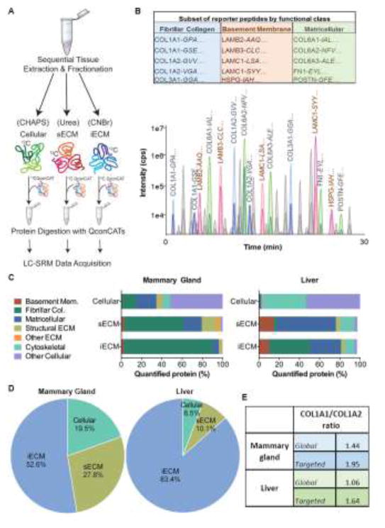
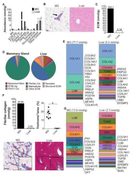
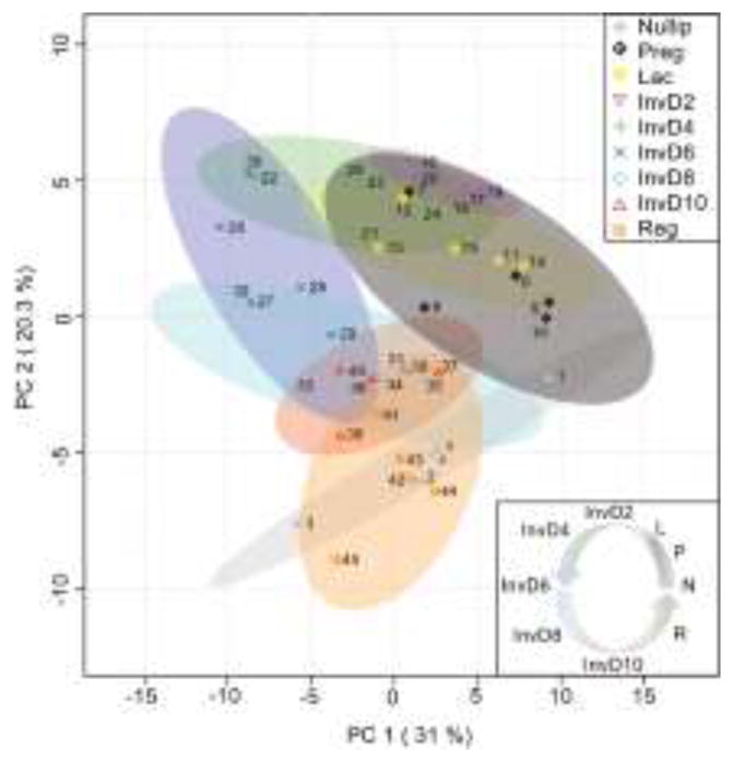
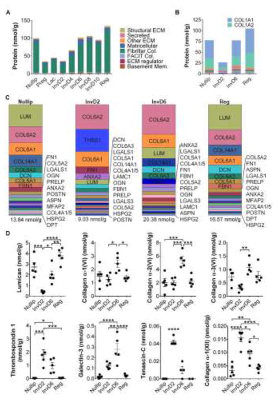
Normal epithelium exists within a dynamic extracellular matrix (ECM) that is tuned to regulate tissue specific epithelial cell function. As such, ECM contributes to tissue homeostasis, differentiation, and disease, including cancer. Though it is now recognized that the functional unit of normal and transformed epithelium is the epithelial cell and its adjacent ECM, we lack a basic understanding of tissue-specific ECM composition and abundance, as well as how physiologic changes in ECM impact cancer risk and outcomes. While traditional proteomic techniques have advanced to robustly identify ECM proteins within tissues, methods to determine absolute abundance have lagged. Here, with a focus on tissues relevant to breast cancer, we utilize mass spectrometry methods optimized for absolute quantitative ECM analysis. Employing an extensive protein extraction and digestion method, combined with stable isotope labeled Quantitative conCATamer (QconCAT) peptides that serve as internal standards for absolute quantification of protein, we quantify 98 ECM, ECM-associated, and cellular proteins in a single analytical run. In rodent models, we applied this approach to the primary site of breast cancer, the normal mammary gland, as well as a common and particularly deadly site of breast cancer metastasis, the liver. We find that mammary gland and liver have distinct ECM abundance and relative composition. Further, we show mammary gland ECM abundance and relative compositions differ across the reproductive cycle, with the most dramatic changes occurring during the pro-tumorigenic window of weaning-induced involution. Combined, this work suggests ECM candidates for investigation of breast cancer progression and metastasis, particularly in postpartum breast cancers that are characterized by high metastatic rates. Finally, we suggest that with use of absolute quantitative ECM proteomics to characterize tissues of interest, it will be possible to reconstruct more relevant in vitro models to investigate tumor-ECM dynamics at higher resolution.
Keywords: Breast cancer; Extracellular matrix; Liver; Liver metastasis; Mammary gland; Mass spectrometry proteomics.
Copyright © 2016 The Authors. Published by Elsevier Ltd.. All rights reserved.
Figures
Similar articles
-
Non-steroidal anti-inflammatory drugs target the pro-tumorigenic extracellular matrix of the postpartum mammary gland.Int J Dev Biol. 2011;55(7-9):745-55. doi: 10.1387/ijdb.113379jo. Int J Dev Biol. 2011. PMID: 22161831
-
Rat mammary extracellular matrix composition and response to ibuprofen treatment during postpartum involution by differential GeLC-MS/MS analysis.J Proteome Res. 2012 Oct 5;11(10):4894-905. doi: 10.1021/pr3003744. Epub 2012 Aug 30. J Proteome Res. 2012. PMID: 22897585 Free PMC article.
-
Mammary ECM composition and function are altered by reproductive state.Mol Carcinog. 2004 Dec;41(4):207-20. doi: 10.1002/mc.20058. Mol Carcinog. 2004. PMID: 15468292
-
Extracellular matrix components in breast cancer progression and metastasis.Breast. 2013 Aug;22 Suppl 2:S66-72. doi: 10.1016/j.breast.2013.07.012. Breast. 2013. PMID: 24074795 Review.
-
Extracellular matrix composition reveals complex and dynamic stromal-epithelial interactions in the mammary gland.J Mammary Gland Biol Neoplasia. 2010 Sep;15(3):301-18. doi: 10.1007/s10911-010-9189-6. Epub 2010 Sep 2. J Mammary Gland Biol Neoplasia. 2010. PMID: 20811805 Review.
Cited by
-
Mammary extracellular matrix directs differentiation of testicular and embryonic stem cells to form functional mammary glands in vivo.Sci Rep. 2017 Jan 10;7:40196. doi: 10.1038/srep40196. Sci Rep. 2017. PMID: 28071703 Free PMC article.
-
Postpartum breast cancer has a distinct molecular profile that predicts poor outcomes.Nat Commun. 2021 Nov 3;12(1):6341. doi: 10.1038/s41467-021-26505-3. Nat Commun. 2021. PMID: 34732713 Free PMC article.
-
Mammary Gland Involution Provides a Unique Model to Study the TGF-β Cancer Paradox.J Clin Med. 2017 Jan 13;6(1):10. doi: 10.3390/jcm6010010. J Clin Med. 2017. PMID: 28098775 Free PMC article. Review.
-
Characterization of weaning-induced breast involution in women: implications for young women's breast cancer.NPJ Breast Cancer. 2020 Oct 16;6:55. doi: 10.1038/s41523-020-00196-3. eCollection 2020. NPJ Breast Cancer. 2020. PMID: 33083533 Free PMC article.
-
Ibuprofen supports macrophage differentiation, T cell recruitment, and tumor suppression in a model of postpartum breast cancer.J Immunother Cancer. 2018 Oct 1;6(1):98. doi: 10.1186/s40425-018-0406-y. J Immunother Cancer. 2018. PMID: 30285905 Free PMC article.
References
-
- Aggeler J, Park CS, Bissell MJ. Regulation of milk protein and basement membrane gene expression: the influence of the extracellular matrix. Journal of dairy science. 1988;71:2830–2842. - PubMed
-
- Schedin P, Mitrenga T, McDaniel S, Kaeck M. Mammary ECM composition and function are altered by reproductive state. Mol Carcinog. 2004;41:207–220. - PubMed
Publication types
MeSH terms
Grants and funding
LinkOut - more resources
Full Text Sources
Other Literature Sources
Molecular Biology Databases
Research Materials
