. 2017 Jun;21(5):1871-1881.
doi: 10.1007/s00784-016-1980-3. Epub 2016 Oct 22.

The mycobiome of root canal infections is correlated to the bacteriome

Affiliations

The mycobiome of root canal infections is correlated to the bacteriome

Ilona F Persoon et al. Clin Oral Investig. 2017 Jun.

Abstract

Objectives: Bacterial infection of the root canal system causes apical periodontitis. Less is known about the role of fungi in these infections. This study aimed to assess the fungal prevalence, abundance, and diversity of root canal infections, as well as the relation between fungi and bacteria present in different parts of the root canal.

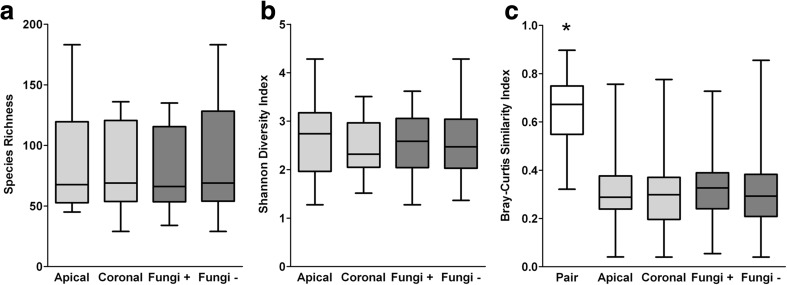
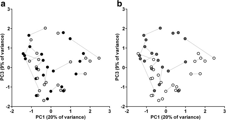
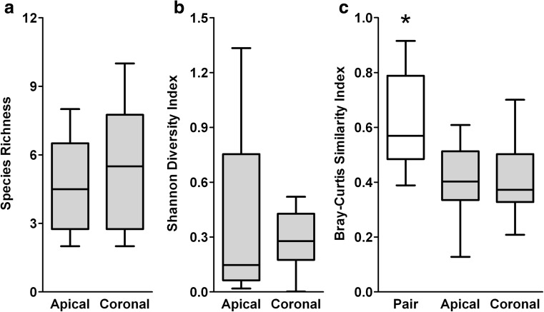
Materials and methods: Twenty-six teeth with primary apical periodontitis were extracted, split in apical and coronal root segments, and cryo-pulverized. Bacteriome profiles of 23 teeth were analyzed based on the V3-V4 hypervariable region of the 16S ribosomal RNA gene. Mycobiome profiles of six teeth were analyzed based on the internal transcribed spacer (ITS) 1 or ITS2 region. Samples were sequenced on the Illumina MiSeq platform.

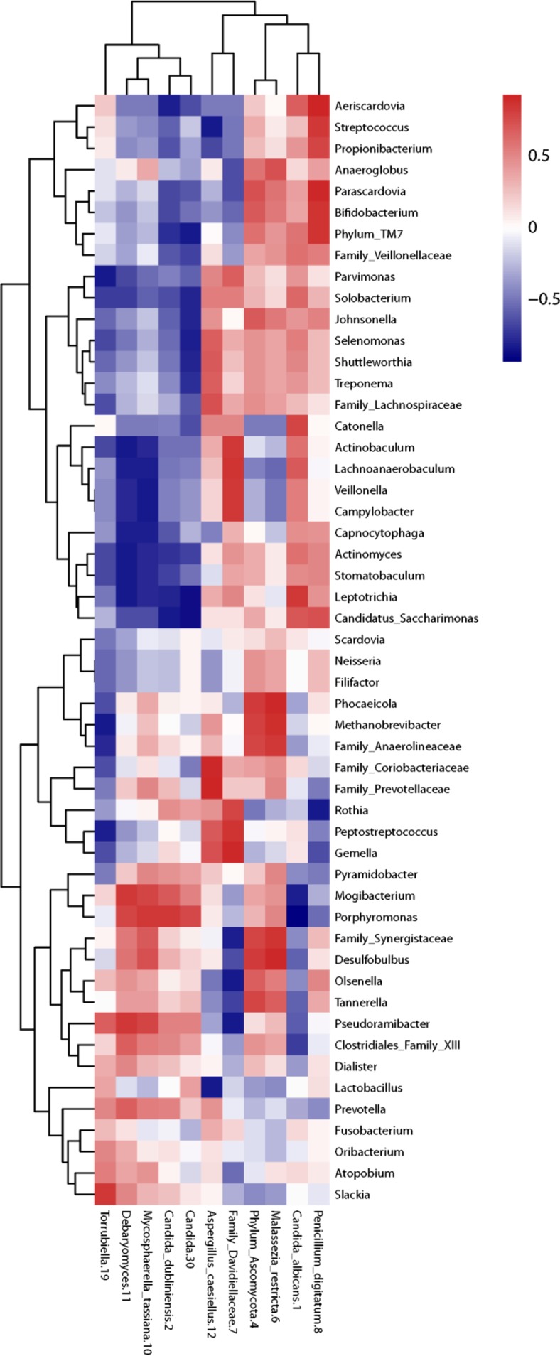
Results: A total of 338 bacterial operational taxonomic units (OTUs), 28 ITS1 OTUs, and 24 ITS2 OTUs were identified. Candida and Malassezia were the most frequently identified fungi. No differences could be found between the bacteriome and mycobiome profiles of the apical and coronal root segments. The bacteriome of fungi-positive root segments contained more Actinomyces, Bifidobacterium, four different Lactobacillus OTUs, Propionibacterium, and Streptococcus. A Spearman correlation matrix between bacteriomes and mycobiomes identified no correlations, but separate clusters could be observed.

Conclusions: A considerable proportion of the root canal infections contain fungi, although fungal diversity is limited. However, when fungi are present, the composition of the bacteriome is clearly different.

Clinical relevance: Interaction between bacteria and fungi in root canal infections may complicate the infection and require alternative treatment strategies.

Keywords: Bacteria; Fungi; Microbiome; Next-generation sequencing; Primary endodontic infection.


Conflict of interest statement

Conflict of interest

The authors declare that they have no conflict of interest.

Funding

This study was funded by the authors and their institutions.

Ethical approval

This article does not contain any studies with animals performed by any of the authors. All procedures performed in studies involving human participants were in accordance with the ethical standards of the institutional and with the 1964 Helsinki declaration and its later amendments.

Informed consent

Informed consent was obtained from all individual participants included in the study.

Figures

Fig. 1
Fig. 1
Diversity analyses of the bacteriome of root canal infections. a Observed species richness (number of OTUs/sample). b Shannon diversity index of within-sample diversity. c Bray-Curtis similarity index of between-sample diversity. For analysis of the species richness and Shannon diversity index, Wilcoxon signed-rank test compared apical (N = 22) to coronal root segments (N = 22; light gray) and the Mann-Whitney U test compared fungi positive (N = 21) to fungi negative root segments (N = 24; dark gray). For analysis of the Bray-Curtis similarity index, the Kruskal-Wallis test compared the paired root samples to the apical and coronal root segments as well as to the fungi-positive and fungi-negative root segments, with post hoc analysis using the Mann-Whitney U test and an FDR correction (q* = 0.033). *p < 0.001
Fig. 2
Fig. 2
A two-dimensional ordination by principal component analysis (PCA) of the bacteriomes. a The bacterial composition of apical (N = 22; light gray) and coronal root segments (N = 22; black) were not statistically significantly different (p = 0.899, one-way permutational multivariate analysis of variance; PERMANOVA). b The bacterial composition of fungi-positive (N = 20; dark gray) and fungi-negative root segments (N = 24; white) was statistically significantly different (p = 0.008; PERMANOVA). Principal components PC1, PC2 (not shown), and PC3 explained 20, 16, and 9 % of the overall variance among samples, respectively. Corresponding apical and coronal root segments are connected with a line
Fig. 3
Fig. 3
Diversity analyses of the mycobiomes of root canal infections as determined through sequencing of the ITS1 region. a Observed species richness (number of OTUs/sample). b Shannon diversity index of within-sample diversity. c Bray-Curtis similarity index of between-sample diversity. Wilcoxon signed-rank test was done to compare apical (N = 6) to coronal root segments (N = 6) for the species richness and Shannon diversity index. The Kruskal-Wallis test was done using the Bray-Curtis similarity indices of the paired root samples to the apical and coronal grouped root segments, with post hoc analysis using the Mann-Whitney U test and an FDR correction (q* = 0.033). *p < 0.001
Fig. 4
Fig. 4
Correlations of the abundances of bacteria and fungi. A Spearman correlation matrix was drafted of bacterial genera prevalent in more than 25 % of the root pairs and at an abundance of more than 0.05 % in correlation with fungal OTUs prevalent in more than one root pair (N = 6). No correlations were significant after FDR correction. Red indicates a positive correlation; blue indicates a negative correlation

References

    1. Kakehashi S, Stanley HR, Fitzgerald RJ. The effects of surgical exposures of dental pulps in germ-free and conventional laboratory rats. Oral Surg Oral Med Oral Pathol. 1965;20:340–349. doi: 10.1016/0030-4220(65)90166-0. - DOI - PubMed
    1. Matsumiya S, Kitamura M. Histopathological and histobacteriological studies of the relation between the condition of sterilization of the interior of the root canal and the healing process of periapical tissues in experimentally infected root canal treatment. Bull Tokyo Dent Coll. 1960;1:1–19.
    1. Wolf TG, Paqué F, Zeller M, et al. Root canal morphology and configuration of 118 mandibular first molars by means of micro–computed tomography: an ex vivo study. J Endod. 2016;42:610–614. doi: 10.1016/j.joen.2016.01.004. - DOI - PubMed
    1. Ricucci D, Siqueira JF., Jr Biofilms and apical periodontitis: study of prevalence and association with clinical and histopathologic findings. J Endod. 2010;36:1277–1288. doi: 10.1016/j.joen.2010.04.007. - DOI - PubMed
    1. Sundqvist G, Figdor D, Persson S, et al. Microbiologic analysis of teeth with failed endodontic treatment and the outcome of conservative re-treatment. Oral Surg Oral Med Oral Pathol Oral Radiol Endod. 1998;85:86–93. doi: 10.1016/S1079-2104(98)90404-8. - DOI - PubMed

LinkOut - more resources