. 2016 Dec:57:282-290.
doi: 10.1016/j.neuro.2016.10.011. Epub 2016 Oct 20.

MDMA decreases glutamic acid decarboxylase (GAD) 67-immunoreactive neurons in the hippocampus and increases seizure susceptibility: Role for glutamate

Affiliations

MDMA decreases glutamic acid decarboxylase (GAD) 67-immunoreactive neurons in the hippocampus and increases seizure susceptibility: Role for glutamate

Courtney L Huff et al. Neurotoxicology. 2016 Dec.

Abstract

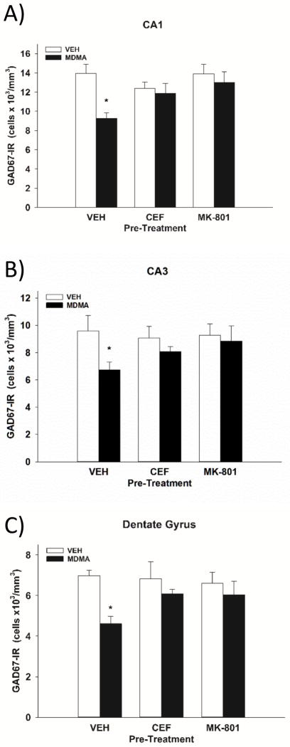
3,4-Methylenedioxy-methamphetamine (MDMA) is a unique psychostimulant that continues to be a popular drug of abuse. It has been well documented that MDMA reduces markers of 5-HT axon terminals in rodents, as well as humans. A loss of parvalbumin-immunoreactive (IR) interneurons in the hippocampus following MDMA treatment has only been documented recently. In the present study, we tested the hypothesis that MDMA reduces glutamic acid decarboxylase (GAD) 67-IR, another biochemical marker of GABA neurons, in the hippocampus and that this reduction in GAD67-IR neurons and an accompanying increase in seizure susceptibility involve glutamate receptor activation. Repeated exposure to MDMA (3×10mg/kg, ip) resulted in a reduction of 37-58% of GAD67-IR cells in the dentate gyrus (DG), CA1, and CA3 regions, as well as an increased susceptibility to kainic acid-induced seizures, both of which persisted for at least 30days following MDMA treatment. Administration of the NMDA antagonist MK-801 or the glutamate transporter type 1 (GLT-1) inducer ceftriaxone prevented both the MDMA-induced loss of GAD67-IR neurons and the increased vulnerability to kainic acid-induced seizures. The MDMA-induced increase in the extracellular concentration of glutamate in the hippocampus was significantly diminished in rats treated with ceftriaxone, thereby implicating a glutamatergic mechanism in the neuroprotective effects of ceftriaxone. In summary, the present findings support a role for increased extracellular glutamate and NMDA receptor activation in the MDMA-induced loss of hippocampal GAD67-IR neurons and the subsequent increased susceptibility to evoked seizures.

Keywords: Excitotoxicity; GABA; Glutamate; MDMA.


Conflict of interest statement

The authors declare no competing financial interests.

Figures

Figure 1
Figure 1
MDMA selectively decreases DAB-labeled GAD67-IR in the rat hippocampus. A) Rats were treated with MDMA (1x or 3×10 mg/kg, ip) or vehicle 7 or 30 days prior to sacrifice. GAD67-IR neurons were counted in 6 right and left images of each brain region from 6 rats/treatment group. *Indicates p<0.05 compared to vehicle. B) Representative images of DAB-labeled GAD67 cells in the CA1, CA3, and DG in rats treated with vehicle or MDMA (3×10 mg/kg, ip).
Figure 2
Figure 2
Glutamate mediates MDMA-induced reductions in GAD67-IR. Animals received ceftriaxone (CEF) (200 mg/kg, ip, daily) for 7 days prior to MDMA, MK-801 (0.3mg/kg, sc, 30 min prior to each injection of MDMA) or vehicle prior to either MDMA (3 × 10 mg/kg, ip) or vehicle for a total of six treatment groups. Animals were sacrificed 7 days after treatment with MDMA, and GAD67-IR was assessed using a stereological technique in the CA1 (A), CA3 (B), and DG (C). GAD67-IR neurons were counted in 2 sections in each brain region from 6–8 rats/treatment group. *Indicates p<0.05 compared to the values for vehicle treated rats.
Figure 3
Figure 3
MDMA increases susceptibility to kainic acid-induced seizures. Rats were given kainic acid (8mg/kg, sc) 7 or 30 days following the administration of either vehicle or MDMA (3×10mg/kg, ip). A) The percentage of rats exhibiting stage 3–5 seizures is depicted. N=8–19 rats/group. B) Of the animals that seized in panel A (n=5–17 rats/group), latency to seizure was recorded as the time (min) at which the animal first exhibited a stage 3–5 behavior. *Indicates p<0.05 compared to the values for vehicle treated rats.
Figure 4
Figure 4
MK-801 and ceftriaxone prevent the MDMA-induced increase in seizure susceptibility. Rats were treated with A) MK-801 (0.3mg/kg, sc) or vehicle 30 min prior to each injection of either MDMA (3×10 mg/kg, ip) or vehicle. B) Ceftriaxone (CEF) (200mg/kg, i.p.) or vehicle was given daily for 7 days prior to either MDMA (3 × 10 mg/kg, ip) or vehicle. Kainic acid (8mg/kg, sc) was administered 7 days after MDMA treatment. The percentage of rats exhibiting stage 3–5 seizures is depicted. N=5–10 rats/group. *Indicates (p<0.05) compared to VEH/VEH. #Indicates (p<0.05) compared to VEH/MDMA.
Figure 5
Figure 5
MDMA-induced hyperthermia was maintained in MK-801- and Ceftriaxone- treated animals. Core body temperatures were recorded every 30 min beginning 1 hr prior and ending 7 hr after the first injection of MDMA. A) Rats were treated with either a single daily injection of ceftriaxone (CEF) (200mg/kg, i.p.) or vehicle for 7 days prior to either MDMA (3 × 10 mg/kg, ip) or vehicle. B) Rats were treated with either MK-801 (0.3mg/kg, sc) or vehicle 30 min prior to each injection of either MDMA (3×10 mg/kg, ip) or vehicle. The values for animals that received MDMA in addition to either CEF or MK-801 were not significantly different from the values for MDMA-treated rats (p>0.05). N=6–8 rats/group.
Figure 6
Figure 6
Ceftriaxone prevents the MDMA-induced increase in extracellular glutamate. Extracellular glutamate was determined in the hippocampus in rats treated daily with ceftriaxone (CEF), (200 mg/kg, ip,) or vehicle for 7 days prior to either MDMA (3 × 10 mg/kg, ip, as indicated by the arrows) or vehicle. N=7–11 rats/group. *Indicates values that differ significantly (p<0.05) from the corresponding average baseline values for vehicle treated animals.

Similar articles

Cited by

References

    1. Abad S, Junyent F, Auladell C, Pubill D, Pallas M, Camarasa J, Escubedo E, Camins A. 3,4-methylenedioxymethamphetamine enhances kainic acid convulsive susceptibility. Prog Neuropsychopharmacol Biol Psychiatry. 2014;54:231–242. - PubMed
    1. Anneken JH, Cunningham JI, Collins SA, Yamamoto BK, Gudelsky GA. MDMA increases glutamate release and reduces parvalbumin-positive GABAergic cells in the dorsal hippocampus of the rat: Role of cyclooxygenase. J Neuroimmune Pharmacol. 2013;8:58–65. - PMC - PubMed
    1. Anneken JH, Gudelsky GA. MDMA produces a delayed and sustained increase in the extracellular concentration of glutamate in the rat hippocampus. Neuropharmacology. 2012;63:1022–1027. - PMC - PubMed
    1. Arellano J, Munoz A, Ballesteros-Yanez I, Sola R, DeFelipe J. Histopathology and reorganization of chandelier cells in the human epileptic sclerotic hippocampus. Brain. 2004;127:45–64. - PubMed
    1. Baumann MH, Wang X, Rothman RB. 3,4-methylenedioxymethamphetamine (MDMA) neurotoxicity in rats: A reappraisal of past and present findings. Psychopharmacology (Berl) 2007;189:407–424. - PMC - PubMed

Publication types

MeSH terms