Review
. 2017 Jan 2:227:158-170.
doi: 10.1016/j.virusres.2016.10.010. Epub 2016 Oct 20.

The family Rhabdoviridae: mono- and bipartite negative-sense RNA viruses with diverse genome organization and common evolutionary origins

Affiliations
Review

The family Rhabdoviridae: mono- and bipartite negative-sense RNA viruses with diverse genome organization and common evolutionary origins

Ralf G Dietzgen et al. Virus Res. .

Abstract

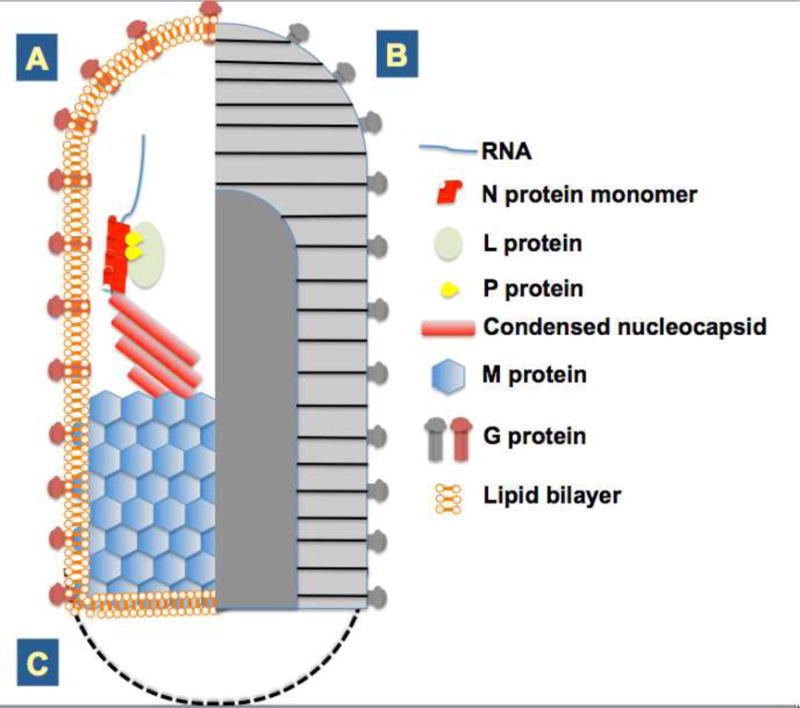
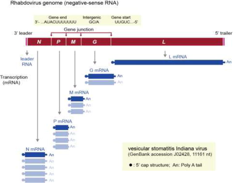
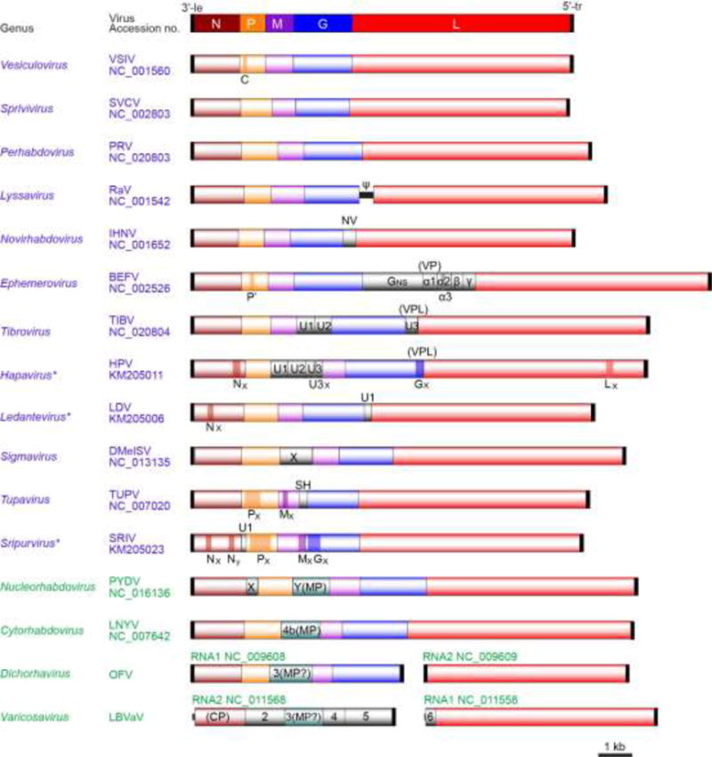
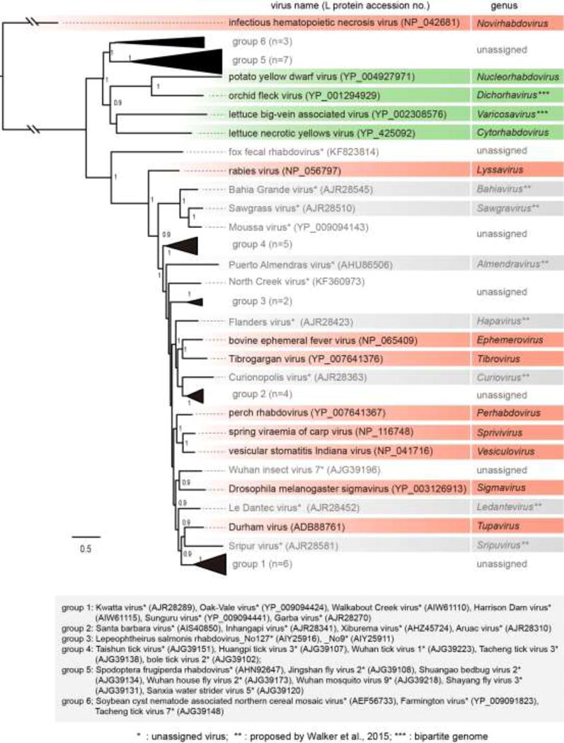
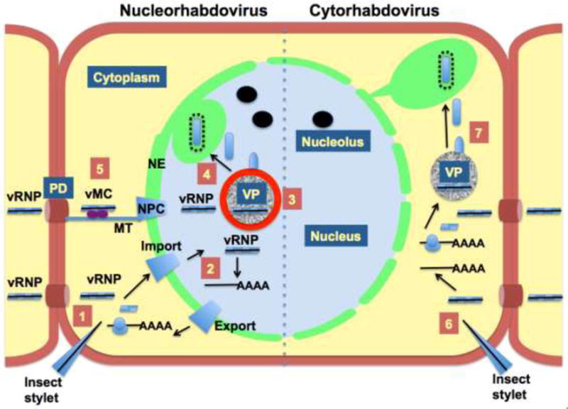
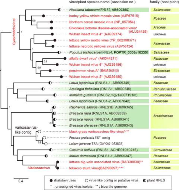
The family Rhabdoviridae consists of mostly enveloped, bullet-shaped or bacilliform viruses with a negative-sense, single-stranded RNA genome that infect vertebrates, invertebrates or plants. This ecological diversity is reflected by the diversity and complexity of their genomes. Five canonical structural protein genes are conserved in all rhabdoviruses, but may be overprinted, overlapped or interspersed with several novel and diverse accessory genes. This review gives an overview of the characteristics and diversity of rhabdoviruses, their taxonomic classification, replication mechanism, properties of classical rhabdoviruses such as rabies virus and rhabdoviruses with complex genomes, rhabdoviruses infecting aquatic species, and plant rhabdoviruses with both mono- and bipartite genomes.

Keywords: Diversity; Genome organization; Negative-sense RNA virus; Replication; Rhabdovirus; Taxonomy.


Figures

Fig. 1
Fig. 1
A. (left half of figure). Schematic representation of rhabdovirus particle internal structure. The single-stranded negative-sense genomic RNA is encapsidated along its entire length by the nucleoprotein N. Associated with the L polymerase and P phosphoprotein, transcriptionally-competent nucleocapsids represent the minimal infectious unit of rhabdoviruses. During virion maturation the nucleocapsid is condensed by the matrix protein, and this complex ultimately buds through host membranes to acquire the lipid envelope and transmembrane glycoprotein present in mature virus particles. B. (right half of figure). External virus particle appearance. Negatively-stained particles appear striated when examined by electron microscopy. Glycoprotein spikes decorate the surface of virions and the central cavity fills with stain which contrasts the space occupied by the matrix-protein condensed nucleocapsid. C. While A and B together show the “rhabdoid” or bullet shape typical of rhabdoviruses adapted to mammalian cells, the virions of plant-adapted rhabdoviruses are typically bacilliform in shape, represented here by a dashed line.
Fig. 2
Fig. 2
Schematic representation of the 3′ to 5′ genome organization (negative-sense) and gene expression of vesicular stomatitis Indiana virus (VSIV, genus Vesiculovirus). VSIV genome encodes the following five canonical rhabdovirus proteins: nucleoprotein (N), phosphoprotein (P), matrix protein (M), glycoprotein (G) and large protein (L, polymerase). The genes are sequentially transcribed presumably through a “stop-start” mechanism, resulting in a 3′–5′ polar gradient of mRNA production (N>P>M >G>L) (see section 3).
Fig. 3
Fig. 3
Comparative genome organization of representative members from sixteen genera of the family Rhabdoviridae (Dietzgen et al., 2011; 2014, Walker et al., 2015). The five canonical structural protein genes (N, P, M, G and L) are shaded in different colors. Other genes including movement protein (MP), viroporin (VP) or viroporin-like protein (VPL) and unknown function protein genes are shaded in grey. Several overlapping and consecutive ORFs (>150 nt) within each transcriptional unit are also shown with dark color (see Walker et al., 2015). The 3′-leader (le) and 5′-trailer (tr) regions are not scaled. Virus names (abbreviation of the member of type species of genera) and Refseq numbers are shown. Asterisks indicate proposed genera.
Fig. 4
Fig. 4
Phylogenetic relationships of members of the family Rhabdoviridae. The tree was constructed by the maximum likelihood (ML) method as described previously (Kondo et al., 2015). The entire L polymerase sequences were aligned using MAFFT 7.0 (Katoh and Toh, 2008) under default settings and ambiguously aligned regions were removed using Gblocks 0.91b (Talavera and Castresana, 2007) with all the options of less stringent selection. A model LG with + I + G + F was selected as the best fit model using PhyML 3.0 (Guindon et al., 2010) with automatic model selection by Smart Model Selection (SMS). The resulting tree was visualized with the FigTree 1.3.1 (http://tree.bio.ed.ac.uk/software/). Numbers at the nodes indicate aLRT values determined using an SH-like calculation (values less than 0.9 are not displayed). Virus names (the member of type species of genera and other selected unclassified rhabdoviruses) and GenBank/Refseq accession numbers (within parentheses) of L proteins are shown. Distantly related rhabdoviruses (group 1 to 6; n, number of sequences) that formed monophyletic clades, probably establishing additional genera in the family, were collapsed into a black triangle. The names and accession numbers of these viruses are shown below the tree.
Fig. 5
Fig. 5
Comparison of the infection cycles of nucleo- and cytorhabdoviruses in plant cells. 1. Nucleorhabdoviruses are initially introduced into plant cells via a mechanical breach of the cell wall by insect vectors or experimentally by abrasion. The introduced viral ribonucleoprotein (vRNP) is imported into nucleus due to interaction with the nuclear import receptor importin-alpha. 2. Within nucleus the vRNP establishes transcription of poly(A)+ transcripts that are exported from the nucleus. Translation of the viral mRNAs in the cytoplasm produces proteins that are imported into nucleus where they aggregate to form the nuclear viroplasms where replication occurs (VP). 3. Nascent nucleocapsids are condensed on the perimeter of the VP, which is enriched in the phosphoprotein (sonchus yellow net virus, SYNV; Goodin, unpublished). 4. Matrix protein condensed nucleocapsids bud into the perinuclear space where mature virions accumulate. In the case of SYNV, the inner membrane of the nuclear envelope (NE) invaginates into the nuclear VP to form virus-containing spherules. 5. Mature virions do not move cell to cell, but may be transmitted to other plants via insect feeding. The vRNP exits the nucleus using the nuclear pore complex (NPC). In the cytoplasm the viral movement complex (vMC) is composed of the vRNP, the viral movement protein, and host factors, which are trafficked towards plasmodesmata (PD) on microtubules (MT), resulting in cell-to-cell spread of the virus. 6. Cytorhabdoviruses are introduced into cells in a manner similar to nucleorhabdoviruses, but carry out their replication cycles in the cytoplasm. 7. Transcription and translation of cytorhabdovirus vRNPs produce the protein pools required to form cytoplasmic VPs for replication. Condensed vRNPs mature in cytoplasmic proliferations on endoplasmic membranes. As above, mature virions do not move from cell to cell, and cytorhabdovirus vRNP are expected to traffic to the PD via a similar vMC mechanism.
Fig. 6
Fig. 6
Phylogenetic relationships of the N (CP) proteins of varicosaviruses, cytorhabdoviruses and rhabdovirus N-like sequences (RLNSs) identified in plant host genomes. The ML tree was constructed using PhyML 3.0 with a best fit model “LG + G”. A part of this analysis was reported by Chiba et al. (2011). The accession numbers of rhabdoviruses and plant RLNSs (RLNS1, 2 and 4) are shown next to the virus or plant species name in the figure (within parentheses). Analyzed sequences were from 13 rhabdoviruses (varicosa- and varicosa-like viruses, cyto- and unclassified rhabdoviruses), 2 varicosavirus-like-contig sequences and 12 plant RNLSs (Chiba et al., 2011). Numbers at the nodes indicate aLRT values (values less than 0.9 are not displayed).

Similar articles

Cited by

References

    1. Abraham G, Banerjee AK. Sequential transcription of the genes of vesicular stomatitis virus. Proc Natl Acad Sci USA. 1976;73:1504–1508. - PMC - PubMed
    1. Adams MJ, et al. Ratification vote on taxonomic proposals to the International Committee on Taxonomy of Viruses. Arch Virol. 2016 doi: 10.1007/s00705-016-2977-6. in press. - DOI - PMC - PubMed
    1. Afonso CL, et al. Taxonomy of the order Mononegavirales – update 2016. Arch Virol. 2016;161:2351–2360. - PMC - PubMed
    1. Ahne W, Bjorklund H, Essbauer S, Fijan N, Kurath G, Winton J. Spring viremia of carp – review. Dis Aquat Organ. 2002;52:261–272. - PubMed
    1. Albertini AA, Wernimont AK, Muziol T, Ravelli RB, Clapier CR, Schoehn G, Weissenhorn W, Ruigrok RW. Crystal structure of the rabies virus nucleoprotein-RNA complex. Science. 2006;313:360–363. - PubMed

Publication types