Review
. 2016 Oct 12:9:47-71.
doi: 10.4137/CMED.S38201. eCollection 2016.

Multiple Effects of Growth Hormone in the Body: Is it Really the Hormone for Growth?

Affiliations
Review

Multiple Effects of Growth Hormone in the Body: Is it Really the Hormone for Growth?

Jesús Devesa et al. Clin Med Insights Endocrinol Diabetes. .

Abstract

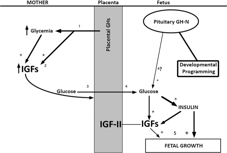
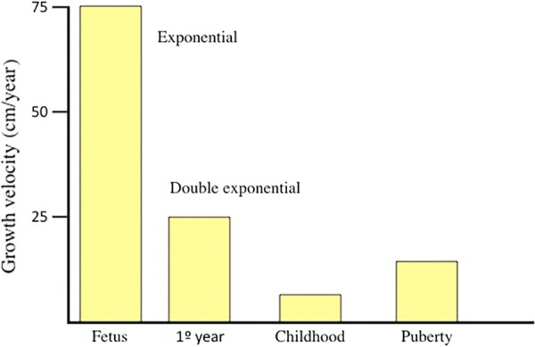
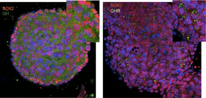
In this review, we analyze the effects of growth hormone on a number of tissues and organs and its putative role in the longitudinal growth of an organism. We conclude that the hormone plays a very important role in maintaining the homogeneity of tissues and organs during the normal development of the human body or after an injury. Its effects on growth do not seem to take place during the fetal period or during the early infancy and are mediated by insulin-like growth factor I (IGF-I) during childhood and puberty. In turn, IGF-I transcription is dependent on an adequate GH secretion, and in many tissues, it occurs independent of GH. We propose that GH may be a prohormone, rather than a hormone, since in many tissues and organs, it is proteolytically cleaved in a tissue-specific manner giving origin to shorter GH forms whose activity is still unknown.

Keywords: cardiovascular system; gonads; growth; growth hormone; liver; nervous system.


Conflict of interest statement

Authors disclose no potential conflicts of interest.

Figures

Figure 1
Figure 1
The products of expression of the hGH gene family share 95% of homology among them. The main GH form corresponds to the 22 kDa GH-N, expressed not only at the pituitary level but also in many different peripheral cells and tissues. About 10% of pituitary GH-N transcripts suffer a physiological postranscriptional modification, occurred by alternative splicing of exon III, giving origin to a 20 kDa variant. Among placental GH genes, the most important seems to be the GH-V, which differs from 22 kDa GH-N in only eight amino acids.
Figure 2
Figure 2
Possible modulation of the hepatic expression of IGF-I gene. GH and insulin may directly induce the transcription of IGF-I gene, but for that, glucose uptake and metabolization by the liver is needed. Hepatic glucose metabolism may directly induce the transcription of IGF-I gene. In addition, liver uptake of some amino acids, induced by GH or insulin, facilitates IGF-I expression, because these amino acids stabilize the IGF-I mRNA.
Figure 3
Figure 3
Preimplantation period. (1) After interacting with GH, most likely secreted from the oviduct, the GHR is translocated to the nucleus of the zygote, promoting the transcription of genes involved in cells proliferation. GH induces the expression of the glucose transporter Glut 1, which leads to glucose uptake by the zygote therefore providing the substrate needed for the production of the energy necessary for beginning proliferation processes shown in (2) It is unclear if IGF-I is secreted from the oviduct in 1 or 2. (3) While the newly formed embryo is progressing along the oviduct to the uterus, there is a clear secretion of GH and IGF-I from the oviduct walls; IGF-I receptor appears now together with GHR in the membrane of cells constituting the morula, facilitating cell proliferation. (4) In a further step, the blastocyst is formed and their cells begin to express GH, GHR, IGF-I, and IGF-I R. This precedes the implantation in the uterus.
Figure 4
Figure 4
Evolution of maternal plasma levels of GH (N and V) and IGF-I during pregnancy. As pregnancy progresses, placental GH-V increases in the maternal blood. This leads to increased IGF-I levels and decreased GH-N levels. The decrease of plasma GH-N is due to the negative feedback that GH-V and IGF-I directly exert on pituitary GH-N secretion and also due to the fact that both GH-V and IGF-I stimulate hypothalamic somatostatin secretion that, in turn, negatively controls pituitary GH-N release. The maternal increase in plasma levels of IGF-II is not shown in the figure. Red arrows indicate negative control.
Figure 5
Figure 5
Fetal growth. (1) Placental GHs, particularly GH-V, are progressively increasing in maternal blood inducing an increase in plasma levels of glucose. This, and presumably (2) the effect of GH-V on liver, increases the hepatic production of IGFs, which optimizes the supply of nutrients to the fetus (3), particularly glucose. The resulting increase in fetal glycemia (4) induces increased release of fetal pancreatic insulin, and the hepatic production of IGFs, which also can be induced by insulin. Despite pituitary fetal GH-N secretion, it is unlikely (4?) that it contributes to fetal hyperglycemia. For its part, the placenta significantly contributes to the increase in fetal plasma concentrations of IGFs by producing IGF-II. Fetal GH most likely plays a role on the developmental programming of virtually all tissues and organs. (5) Insulin, mainly, and IGFs are the hormones that the fetus needs for growing. Abbreviation: +, stimulates.
Figure 6
Figure 6
Growth velocity along life. The maximum growth velocity is observed during the fetal life, reaching 75 cm/year and following an exponential curve. After birth, growth velocity follows a double exponential curve; the consequence is that the high growth velocity observed after birth progressively slows since 6–10 months of age, indicating that two dfferent kinds of factors are acting in this period. During childhood, growth velocity (lineal now) decreases to 5 cm/year, and when puberty begins, a number of factors (mainly sex steroids) acting on pituitary GH synthesis and release again increase growth velocity until 10–12 cm/year.
Figure 7
Figure 7
Hepatic expression of GH. GH mRNA from the pituitary and liver (partially hepatectomized rats) was retrotranscripted with specific primers and the resultant cDNAs were resolved in 2% agarose and stained with ethidium bromide, before and after using the enzyme of restriction RsaI. As the figure shows pituitary (pit) and liver (liv), GH was detected with the expected molecular weight: 328 bp, because the primers used flanked a region situated between exons 4 and 5 of rat GH gene. The bands obtained after cutting the main GH amplified with RsaI led to the appearance of two bands in the molecular weight expected (238 and 90 bp), both in pituitary and liver GH. Black arrows indicate the main GH product. Blue arrows indicate the products obtained after cutting with RsaI. Reprinted with permission of the publisher. Source: J Devesa, Devesa P, Reimunde P. Growth hormone: actions and preventive and therapeutic applications. Med Clin (Barc) 2010; 135 (14): 665-670. Copyright © 2009 Elsevier Spain, S.L. All rights reserved. Abbreviations: MWM, molecular weight markers; bp, basepairs.
Figure 8
Figure 8
Adrenal expression of GH in rats. GH mRNA from an adrenal removed (control) and from the contralateral adrenal gland (24 hours after unilateral adrenalectomy) was retrotranscripted with specific primers, and the resultant cDNAs were resolved in 2% agarose and stained with ethidium bromide. GH products were detected in the expected molecular weight: 328 bp. Adrenalectomy markedly increased the expression of GH in the contralateral gland 24 hours after the adrenalectomy (Ax 24 h). This may indicate that, apart from ACTH action, GH is overexpressed in the adrenal gland for mediating the compensatory hypertrophy. Reprinted with permission of the publisher. Source: J Devesa, Devesa P, Reimunde P. Growth hormone: actions and preventive and therapeutic applications. Med Clin (Barc) 2010; 135 (14): 665-670. Copyright © 2009 Elsevier Spain, S.L. All rights reserved. Abbreviation: MWM, molecular weight markers.
Figure 9
Figure 9
GH and GHR are expressed in NSCs from mice. NSCs obtained from nine-day-old mice cultured in proliferation media, free of GH, form neurospheres in which confocal microscopy immunoreactivity for SOX2 in the nuclei indicate that these cells are in an early phase of self-renewal. These cells show high immunoreactivity (green labeling) for GH (left) and its receptor (right), indicating that both GH and GHR are expressed in NSCs. Magnification: 40×. © 2012 Devesa J, Devesa P, Reimunde P, Arce V. Published in Growth Hormone and Kynesitherapy for Brain Injury Recovery under CC BY 3.0 license. Available from: http://dx.doi.org/10.5772/26998. Abbreviation: NSCs, neural stem cells.

Similar articles

Cited by

References

    1. Evans HM, Long JA. The effect of the anterior lobe administered intraperitoneally upon growth maturation, and oestrus cycles of the rat. Anatomical Record. 1921;21:62–63.
    1. Raben MS. Treatment of a pituitary dwarf with human growth hormone. J Clin Endocrinol Metab. 1958;18:901–903. - PubMed
    1. Keohane C. Prion diseases in men. Arch Anat Cytol Pathol. 1994;42:69–75. - PubMed
    1. Stockdale T. Contaminated material caused Creutzfeld-Jakob disease (CJD) in some undersized children who were treated with growth hormone (GH) Nutr Health. 2000;14:141–142. - PubMed
    1. Sairam MR, Chrétien M, Li CH. On the isolation of human pituitary hormones. J Clin Endocrinol Metab. 1978;47:1002–1008. - PubMed

LinkOut - more resources