. 2016 Oct 7:7:457.
doi: 10.3389/fphys.2016.00457. eCollection 2016.

Characterization of a Mouse Model of Emphysema Induced by Multiple Instillations of Low-Dose Elastase

Affiliations

Characterization of a Mouse Model of Emphysema Induced by Multiple Instillations of Low-Dose Elastase

Milena V Oliveira et al. Front Physiol. .

Abstract

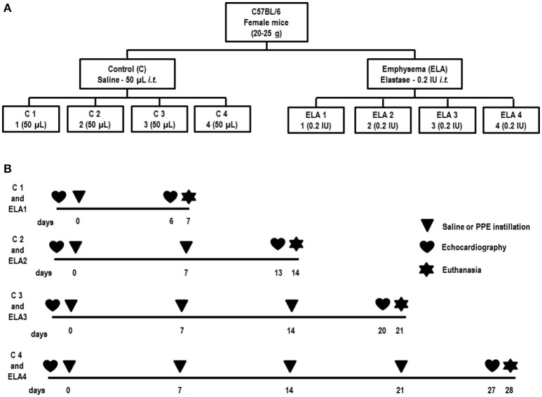
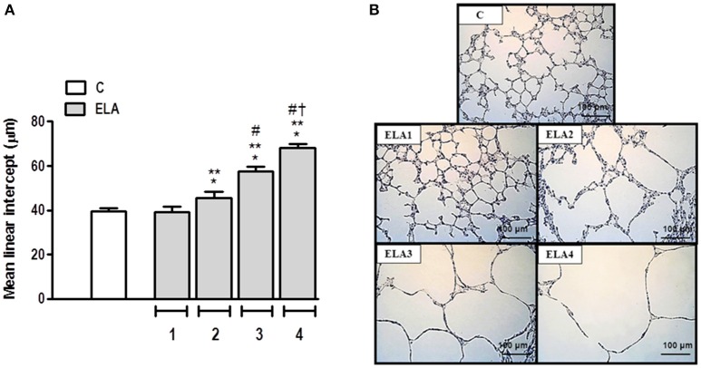
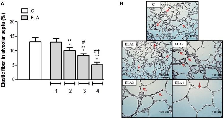
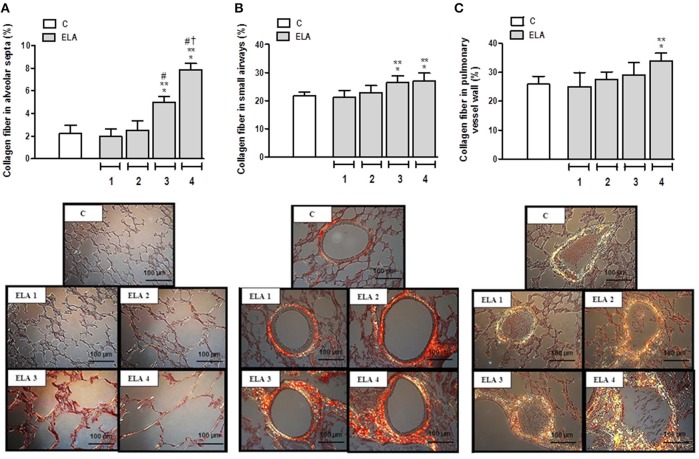
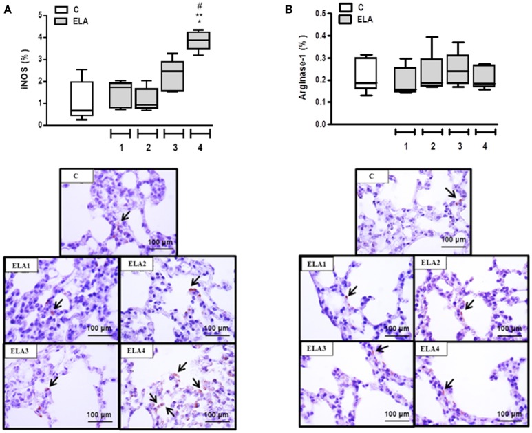
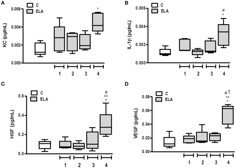
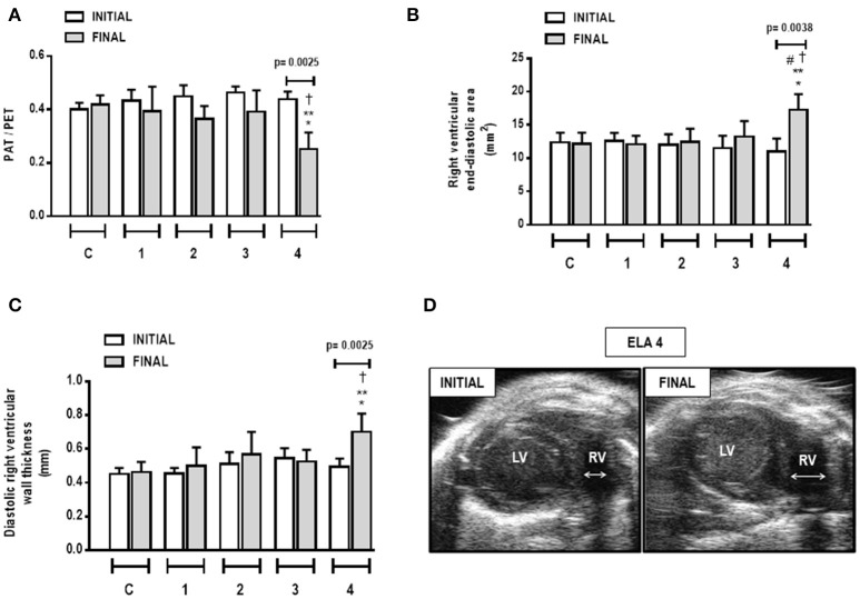
Many experimental models have been proposed to study the pathophysiological features of emphysema, as well as to search for new therapeutic approaches for acute or chronically injured lung parenchyma. We aimed to characterize an emphysema model induced by multiple instillations of elastase by tracking changes in inflammation, remodeling, and cardiac function after each instillation. Forty-eight C57BL/6 mice were randomly assigned across two groups. Emphysema (ELA) animals received 1, 2, 3, or 4 intratracheal instillations of pancreatic porcine elastase (PPE, 0.2 IU) with a 1-week interval between them. Controls (C) received saline following the same protocol. Before and after implementation of the protocol, animals underwent echocardiographic analysis. After the first instillation of PPE, the percentage of mononuclear cells in the lung parenchyma increased compared to C (p = 0.0001). The second instillation resulted in hyperinflated alveoli, increased mean linear intercept, and reduced elastic fiber content in lung parenchyma compared to C (p = 0.0197). Following the third instillation, neutrophils and collagen fiber content in alveolar septa and airways increased, whereas static lung elastance was reduced compared to C (p = 0.0094). After the fourth instillation, the percentage of M1 macrophages in lungs; levels of interleukin-1β (IL-1β), keratinocyte-derived chemokine, hepatocyte growth factor (HGF), and vascular endothelial growth factor (VEGF); and collagen fiber content in the pulmonary vessel wall were increased compared to C (p = 0.0096). At this time point, pulmonary arterial hypertension was apparent, with increased diastolic right ventricular wall thickness. In conclusion, the initial phase of emphysema was characterized by lung inflammation with predominance of mononuclear cells, whereas at the late stage, impairment of pulmonary and cardiovascular functions was observed. This model enables analysis of therapies at different time points during controlled progression of emphysema. Accordingly, early interventions could focus on the inflammatory process, while late interventions should focus on restoring cardiorespiratory function.

Keywords: cardiac function; collagen; elastance; elastic fiber; emphysema; inflammation.


Figures

Figure 1
Figure 1
Schematic flow chart (A) and timeline of study design (B). C group, control (animals that received 1, 2, 3, or 4 intratracheal instillations of saline at 1-week intervals). ELA1, single intratracheal instillation of pancreatic porcine elastase (PPE). ELA2, two instillations of PPE given 1 week apart. ELA3, three instillations of PPE at 1-week intervals. ELA4, four instillations of PPE at 1-week intervals.
Figure 2
Figure 2
Mean linear intercept (A) and representative photomicrographs of lung parenchyma stained with hematoxylin–eosin (H&E) (B). C group, control (animals that received 1, 2, 3, or 4 intratracheal instillations of saline at 1-week intervals). ELA1, single intratracheal instillation of pancreatic porcine elastase (PPE). ELA2, two instillations of PPE given 1 week apart. ELA3, three instillations of PPE at 1-week intervals. ELA4, four instillations of PPE at 1-week intervals. Values are means + SD of six animals in each group. *Significantly different from C group (p < 0.05). **Significantly different from ELA1 group (p < 0.05). #Significantly different from ELA2 group (p < 0.05). Significantly different from ELA3 group (p < 0.05).
Figure 3
Figure 3
Elastic fiber content in alveolar septa (A) and representative photomicrographs of lung parenchyma stained with Weigert's resorcin fuchsin method with oxidation (elastic fibers) (B). Arrows: Elastic fibers (stained black). C group, control (animals that received 1, 2, 3, or 4 intratracheal instillations of saline at 1-week intervals). ELA1, single intratracheal instillation of pancreatic porcine elastase (PPE). ELA2, two instillations of PPE given 1 week apart. ELA3, three instillations of PPE at 1-week intervals. ELA4, four instillations of PPE at 1-week intervals. Values are means + SD of six animals in each group. *Significantly different from C group (p < 0.05). **Significantly different from ELA1 group (p < 0.05). #Significantly different from ELA2 group (p < 0.05). Significantly different from ELA3 group (p < 0.05).
Figure 4
Figure 4
Collagen fiber content and representative photomicrographs of alveolar septa (A), airways (B), and pulmonary vessel wall (C) stained with the Picrosirius-polarization method. C group, control (animals that received 1, 2, 3, or 4 intratracheal instillations of saline at 1-week intervals). ELA1, single intratracheal instillation of pancreatic porcine elastase (PPE). ELA2, two instillations of PPE given 1 week apart. ELA3, three instillations of PPE at 1-week intervals. ELA4, four instillations of PPE at 1-week intervals. Values are means + SD of six animals in each group. *Significantly different from C group (p < 0.05). **Significantly different from ELA1 group (p < 0.05). #Significantly different from ELA2 group (p < 0.05). Significantly different from ELA3 group (p < 0.05).
Figure 5
Figure 5
Immunohistochemistry for iNOS (A) and arginase-1 (B). Note positive cells stained brown (arrows). C group, control (animals that received 1, 2, 3, or 4 intratracheal instillations of saline at 1-week intervals). ELA1, single intratracheal instillation of pancreatic porcine elastase (PPE). ELA2, two instillations of PPE given 1 week apart. ELA3, three instillations of PPE at 1-week intervals. ELA4, four instillations of PPE at 1-week intervals. Values are shown as box-plots (median, interquartile range, minimum, and maximum) of six animals in each group. *Significantly different from C group (p < 0.05). **Significantly different from ELA1 group (p < 0.05). #Significantly different from ELA2 group (p < 0.05).
Figure 6
Figure 6
Levels of keratinocyte-derived chemokine (KC, a mouse analog of interleukin-8) (A), interleukin (IL)-1β (B), hepatocyte growth factor (HGF) (C), and vascular endothelial growth factor (VEGF) (D) in lung tissue. C group, control (animals that received 1, 2, 3, or 4 intratracheal instillations of saline at 1-week intervals). ELA1, single intratracheal instillation of pancreatic porcine elastase (PPE). ELA2, two instillations of PPE given 1 week apart. ELA3, three instillations of PPE at 1-week intervals. ELA4, four instillations of PPE at 1-week intervals. Values are shown as box-plots (median, interquartile range, minimum, and maximum) of six animals in each group. *Significantly different from C group (p < 0.05). **Significantly different from ELA1 group (p < 0.05). #Significantly different from ELA2 group (p < 0.05). Significantly different from ELA3 group (p < 0.05).
Figure 7
Figure 7
Pulmonary artery acceleration time/pulmonary artery ejection time (PAT/PET) ratio (A), right ventricular end-diastolic area (B), diastolic right ventricular wall thickness (C), and short-axis B-dimensional views of both ventricles in ELA4 animals (D). LV, left ventricle; RV, right ventricle; INITIAL, before instillation of saline or PPE; FINAL, after instillation of saline or PPE. Values are means + SD of six animals in each group. *Significantly different from C group (p < 0.05). **Significantly different from ELA1-FINAL group (p < 0.05). #Significantly different from ELA2-FINAL group (p < 0.05). Significantly different from ELA3-FINAL group (p < 0.05).

Similar articles

Cited by

References

    1. Abbas A. E., Franey L. M., Marwick T., Maeder M. T., Kaye D. M., Vlahos A. P., et al. . (2013). Noninvasive assessment of pulmonary vascular resistance by doppler echocardiography. J. Am. Soc. Echocardiogr. 26, 1170–1177. 10.1016/j.echo.2013.06.003 - DOI - PubMed
    1. Agustí A. G., Noguera A., Sauleda J., Sala E., Pons J., Busquets X. (2003). Systemic effects of chronic obstructive pulmonary disease. Eur. Respir. J. 21, 347–360. 10.1183/09031936.03.00405703 - DOI - PubMed
    1. Antunes M. A., Abreu S. C., Cruz F. F., Teixeira A. C., Lopes-Pacheco M., Bandeira E., et al. . (2014). Effects of different mesenchymal stromal cell sources and delivery routes in experimental emphysema. Respir. Res. 15:118. 10.1186/s12931-014-0118-x - DOI - PMC - PubMed
    1. Beard J. T., Newman J. H., Loyd J. E., Byrd B. F. (1991). Doppler estimation of changes in pulmonary artery pressure during hypoxic breathing. J. Am. Soc. Echocardiogr. 4, 121–130. - PubMed
    1. Cheng S. L., Wang H. C., Yu C. J., Tsao P. N., Carmeliet P., Cheng S. J., et al. . (2009). Prevention of elastase-induced emphysema in placenta growth factor knock-out mice. Respir. Res. 10, 115–125. 10.1186/1465-9921-10-115 - DOI - PMC - PubMed

LinkOut - more resources