Review
. 2016 Oct 7:6:122.
doi: 10.3389/fcimb.2016.00122. eCollection 2016.

Experimental Models of Foamy Macrophages and Approaches for Dissecting the Mechanisms of Lipid Accumulation and Consumption during Dormancy and Reactivation of Tuberculosis

Affiliations
Review

Experimental Models of Foamy Macrophages and Approaches for Dissecting the Mechanisms of Lipid Accumulation and Consumption during Dormancy and Reactivation of Tuberculosis

Pierre Santucci et al. Front Cell Infect Microbiol. .

Abstract

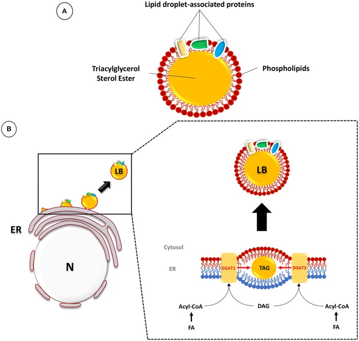
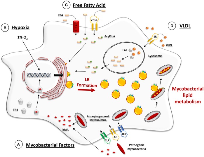
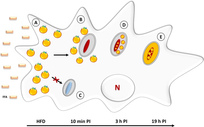
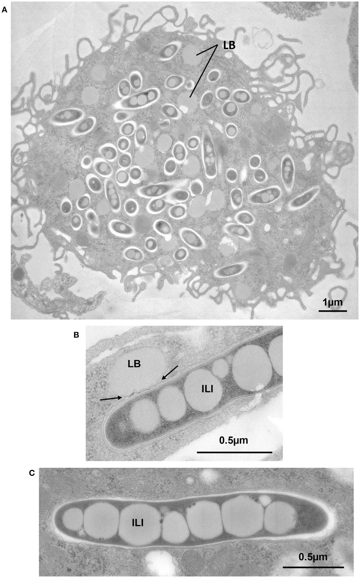
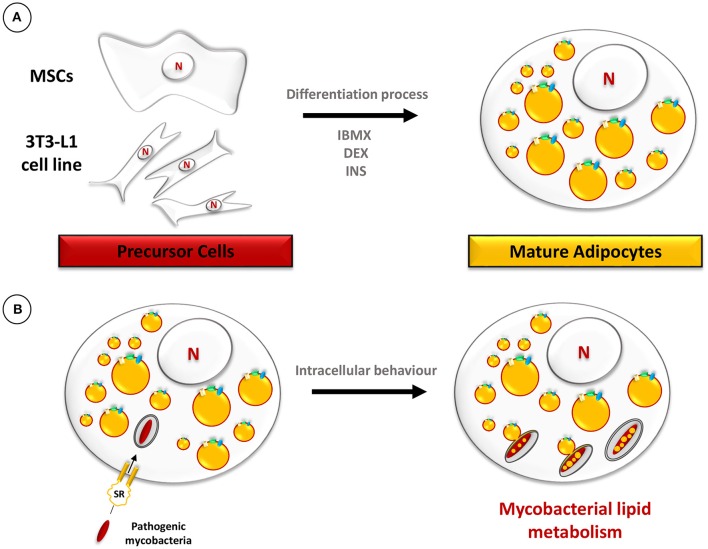
Despite a slight decline since 2014, tuberculosis (TB) remains the major deadly infectious disease worldwide with about 1.5 million deaths each year and with about one-third of the population being latently infected with Mycobacterium tuberculosis, the etiologic agent of TB. During primo-infection, the recruitment of immune cells leads to the formation of highly organized granulomas. Among the different cells, one outstanding subpopulation is the foamy macrophage (FM), characterized by the abundance of triacylglycerol-rich lipid bodies (LB). M. tuberculosis can reside in FM, where it acquires, from host LB, the neutral lipids which are subsequently processed and stored by the bacilli in the form of intracytosolic lipid inclusions (ILI). Although host LB can be viewed as a reservoir of nutrients for the pathogen during latency, the molecular mechanisms whereby intraphagosomal mycobacteria interact with LB and assimilate the LB-derived lipids are only beginning to be understood. Past studies have emphasized that these physiological processes are critical to the M. tuberculosis infectious-life cycle, for propagation of the infection, establishment of the dormancy state and reactivation of the disease. In recent years, several animal and cellular models have been developed with the aim of dissecting these complex processes and of determining the nature and contribution of their key players. Herein, we review some of the in vitro and in vivo models which allowed to gain significant insight into lipid accumulation and consumption in M. tuberculosis, two important events that are directly linked to pathogenicity, granuloma formation/maintenance and survival of the tubercle bacillus under non-replicative conditions. We also discuss the advantages and limitations of each model, hoping that this will serve as a guide for future investigations dedicated to persistence and innovative therapeutic approaches against TB.

Keywords: adipocyte; amoeba; granuloma; lipid body; mycobacteria; pathogenesis.


Figures

Figure 1
Figure 1
Composition and biogenesis of lipid bodies (LB) in mammalian cells. (A) The LB is composed of a core of TAG and sterol esters surrounded by a phospholipid monolayer and associated proteins. (B) TAG synthesis occurs between the two membrane leaflets of the endoplasmic reticulum (ER) (inner leaflet in blue and outer leaflet in red). The prevailing model suggests that host LB originate at the ER, where fatty acids (FA) are resynthesized to form triacylglycerol (TAG) and sterol esters. These compounds accumulate in the hydrophobic space between the two leaflets (in red: outer leaflet; in blue: inner leaflet) of the ER bilayer membrane. The final step is catalyzed by the diacylglycerol acyltransferases DGAT1 and DGAT2. The conversion of diacylglycerol (DAG) and Acyl-CoA into TAG leads to the formation of a lipid globule which will expand dynamically and finally be released into the cytosol as a mature LB surrounded by phospholipids of the cytosolic leaflet of the ER bilayer membrane. Adapted from (Murphy, ; Kassan et al., ; Pol et al., 2014).
Figure 2
Figure 2
Ex vivo-induced mechanisms promoting LB formation in macrophage models. (A) Phagocytic uptake of mycobacteria through scavenger (SR), C-type lectin (CLR), and Toll-like receptors (TLR). Lipid-containing moeities of the mycobacterial cell wall, such as keto-mycolic acids (kMA), are actively trafficked out of mycobacterium-containing phagosomes via fission vesicles (Beatty et al., 2000). At some point during or after vesicle trafficking through early and late endosomes, kMA, or their degradation products, are recognized by the testicular-receptor 4 (TR4) transcriptional factor which then translocates inside the nucleus and triggers a transcriptional response that promotes LB formation (Korf et al., ; Mahajan et al., ; Dkhar et al., 2014). (B) Cell culturing under conditions of hypoxia (1% O2) induces several transcriptional changes which act upon lipid metabolism and promote LB formation (Bostrom et al., ; Daniel et al., 2011). (C) Cell exposure to externally added free fatty acids (FFA). Internalization of FFA may occur via three main pathways: (i) diffusion through the plasma membrane, (ii) binding to the fatty acid transporter protein (FATP) or (iii) binding to SR, such as CD36. Once inside the cytosol, FFA are activated by an Acyl-CoA synthase and transported into the ER where LB are formed (Listenberger et al., ; Glatz et al., 2010). (D) Cell exposure to exogenous TAG-enriched VLDL. Lipids from the lipoproteins (VLDL) that are internalized by scavenger receptor-mediated endocytosis undergo hydrolysis in lysosomes by the lysosomal acid lipase (LAL). This provides the FFA for the subsequent building-up of TAG for LB formation, as mentioned above (Shashkin et al., ; Caire-Brändli et al., 2014).
Figure 3
Figure 3
Neutral lipid accumulation in Mycobacterium marinum-infected D. discoideum. (A) Prior to infection, amoebae are incubated with palmitic acid, corresponding to the High Fat Diet (HFD). This triggers the rapid formation of LB which leads to a foamy appearance. (B) Within 10 min post-infection with M. marinum, LB are gathered around the mycobacteria-containing vacuoles. (C) Infection with amikacin-killed mycobacteria fails to induce LB movement/relocation to the mycobacteria-containing vacuoles. (D) At 3 h post-infection, intact LB are found within the lumen of M. marinum—containing vacuoles. At this stage, a few ILI are already detectable inside the bacteria. (E) At later stages (19 h post-infection), neutral lipids are homogenously distributed inside the mycobacterial cytosol where they accumulate in the form of large ILI.
Figure 4
Figure 4
Exposure of BMDM to VLDL induces foam cell formation, transfer of host TAG to mycobacterium-containing phagosomes by fusion of LB with phagosomes and accumulation of TAG in the form of ILI. BMDM were infected with M. avium or M. bovis BCG. At day 6 post-infection., cells were exposed to VLDL for 24 h, fixed and processed for EM. (A) M. avium-infected cell displaying large amounts of LB, typical of FM. (B) BCG-containing phagosome in direct contact with an LB showing deformation of the phagosome membrane (arrows). (C) Intraphagosomal M. avium displaying several large ILI, 0.4 to 0.5 μm in width, and extending across the full width of the M. avium cytoplasm.
Figure 5
Figure 5
Schematic representation of the adipocyte model. (A) The differentiation procedure of precursor cells, such as mesenchymal stem cells (MSCs) or 3T3-L1, into mature adipocytes includes treatment with 3-isobutyl-1-methylxanthine (IBMX), dexamethasone (DEX), and insulin (INS). (B) Mature adipocytes, filled with LB, are infected with pathogenic mycobacteria which are phagocytosed after binding to scavenger receptors (Neyrolles et al., 2006). Ultimately, mycobacteria accumulate lipids in the form of ILI.

Similar articles

Cited by

References

    1. Agarwal P., Khan S. R., Verma S. C., Beg M., Singh K., Mitra K., et al. . (2014). Mycobacterium tuberculosis persistence in various adipose depots of infected mice and the effect of anti-tubercular therapy. Microbes Infect. 16, 571–580. 10.1016/j.micinf.2014.04.006 - DOI - PubMed
    1. Alvarez H. M. (2016). Triacylglycerol and wax ester-accumulating machinery in prokaryotes. Biochimie 120, 28–39. 10.1016/j.biochi.2015.08.016 - DOI - PubMed
    1. Ates L. S., Ummels R., Commandeur S., Van De Weerd R., Sparrius M., Weerdenburg E., et al. . (2015). Essential role of the ESX-5 secretion system in outer membrane permeability of pathogenic Mycobacteria. PLoS Genet. 11:e1005190. 10.1371/journal.pgen.1005190 - DOI - PMC - PubMed
    1. Baek S. H., Li A. H., Sassetti C. M. (2011). Metabolic regulation of mycobacterial growth and antibiotic sensitivity. PLoS Biol. 9:e1001065. 10.1371/journal.pbio.1001065 - DOI - PMC - PubMed
    1. Bakala N'goma J. C., Schué M., Carriere F., Geerlof A., Canaan S. (2010). Evidence for the cytotoxic effects of Mycobacterium tuberculosis phospholipase C towards macrophages. Biochim. Biophys. Acta 1801, 1305–1313. 10.1016/j.bbalip.2010.08.007 - DOI - PubMed

Publication types

LinkOut - more resources