. 2014 Mar 31:2:23-33.
doi: 10.2147/HP.S54404. eCollection 2014.

Hypoxia-inducible factor-mediated induction of WISP-2 contributes to attenuated progression of breast cancer

Affiliations

Hypoxia-inducible factor-mediated induction of WISP-2 contributes to attenuated progression of breast cancer

Jerry H Fuady et al. Hypoxia (Auckl). .

Abstract

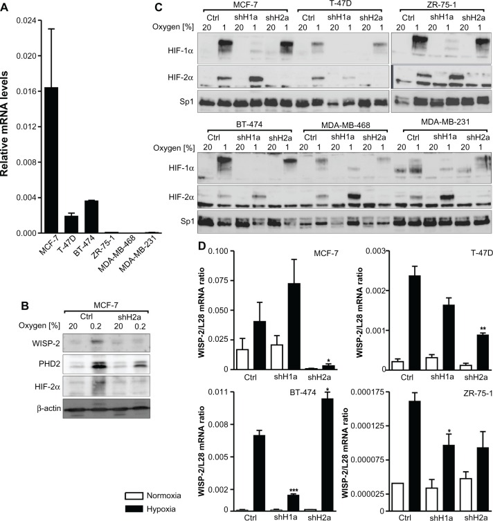
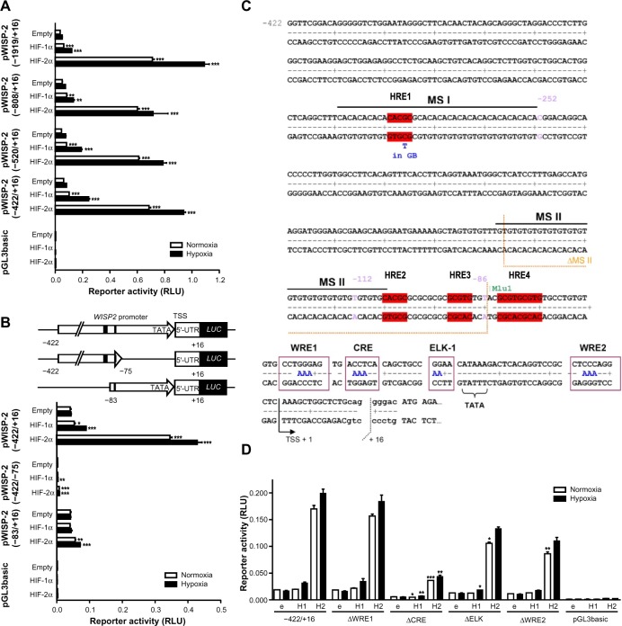
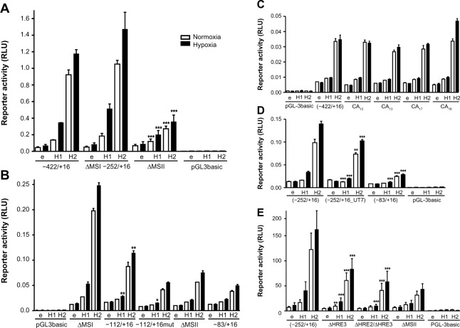
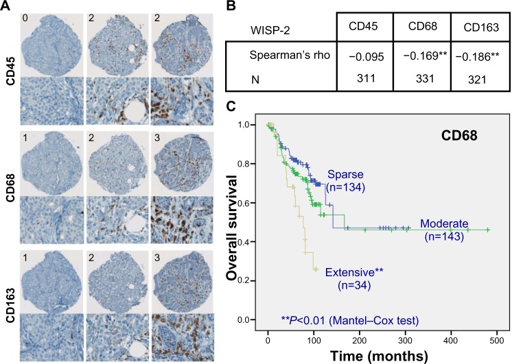
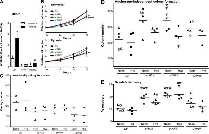
Hypoxia and the hypoxia-inducible factor (HIF) signaling pathway trigger the expression of several genes involved in cancer progression and resistance to therapy. Transcriptionally active HIF-1 and HIF-2 regulate overlapping sets of target genes, and only few HIF-2 specific target genes are known so far. Here we investigated oxygen-regulated expression of Wnt-1 induced signaling protein 2 (WISP-2), which has been reported to attenuate the progression of breast cancer. WISP-2 was hypoxically induced in low-invasive luminal-like breast cancer cell lines at both the messenger RNA and protein levels, mainly in a HIF-2α-dependent manner. HIF-2-driven regulation of the WISP2 promoter in breast cancer cells is almost entirely mediated by two phylogenetically and only partially conserved functional hypoxia response elements located in a microsatellite region upstream of the transcriptional start site. High WISP-2 tumor levels were associated with increased HIF-2α, decreased tumor macrophage density, and a better prognosis. Silencing WISP-2 increased anchorage-independent colony formation and recovery from scratches in confluent cell layers of normally low-invasive MCF-7 cancer cells. Interestingly, these changes in cancer cell aggressiveness could be phenocopied by HIF-2α silencing, suggesting that direct HIF-2-mediated transcriptional induction of WISP-2 gene expression might at least partially explain the association of high HIF-2α tumor levels with prolonged overall survival of patients with breast cancer.

Keywords: invasion; metastasis; motility; oxygen; transcriptional regulation; tumor.


Figures

Figure 1
Figure 1
HIFα isoform-specific regulation of WISP-2 transcription. (A) Basal WISP-2 mRNA levels were determined by reverse transcription quantitative polymerase chain reaction in the six breast cancer cell lines indicated using a calibrated internal standard. Shown are mean values ± the standard error of the mean of three independent experiments. (B) MCF-7 cells were cultured in normoxic conditions or exposed to hypoxia for 24 hours, and analyzed by immunoblotting using antibodies derived against WISP-2, the HIF target prolyl-4-hydroxylase domain 2 (PHD2), HIF-2α, or the constitutively expressed control protein, β-actin. (C) Cells were stably transfected with either control shRNA or shRNAs targeting HIF-1α (shH1a) or HIF-2α (shH2a) and analyzed by immunoblotting of nuclear extracts using antibodies derived against HIF-1α, HIF-2α, or the constitutively expressed control transcription factor, Sp1. (D) Hypoxic WISP-2 mRNA induction was determined in the indicated cell lines which were stably transfected with either control or shRNA constructs as in (C). WISP-2 mRNA levels were quantified by reverse transcription quantitative polymerase chain reaction and normalized to the mRNA levels of the ribosomal protein, L28. Shown are mean values ± the standard errors of the mean of three independent experiments. (D) For statistical evaluation of the hypoxically exposed cells, the effects of HIF-1α or HIF-2α silencing were compared with the control shRNA transfected cells. *P<0.05; **P<0.01; ***P<0.001. Abbreviations: Ctrl, control; HIF, hypoxia-inducible factor; WISP-2, Wnt-1 induced signaling protein 2; mRNA, messenger RNA.
Figure 2
Figure 2
Characterization of the WISP2 promoter. Dual luciferase reporter gene experiments in MCF-7 cells transfected with either HIF-1α or HIF-2α overexpression vectors together with the indicated firefly luciferase vectors and a renilla luciferase control vector, after 24 hours under normoxic or hypoxic (1% O2) conditions. (A) Promoter truncations from positions −1919 to −422 upstream of the transcriptional start site. (B) Potential hypoxia response elements are depicted by black marks (upper panel). HIF responsiveness was lost in the minimal promoter (lower panel). (C) Four potential hypoxia response elements in the WISP2 5′ flanking region are indicated in red. (D) MCF-7 shH2a cells were cotransfected with either the wild-type (−422) promoter construct or constructs bearing the indicated mutations, together with either an empty (e) vector, or a HIF-1α (H1) or HIF-2α (H2) overexpression vector. Shown are mean values ± the standard errors of the mean of three (four for D) independent experiments performed in triplicate. For statistical evaluation, the effects of HIF-1α and HIF-2α overexpression were compared with the respective empty vector controls (A and B), or were first normalized to the empty vector control values and then compared with the respective normalized values of the wild-type −422/+16 promoter construct (D). *P<0.05; **P<0.01; ***P<0.001. Abbreviations: HIF, hypoxia-inducible factor; WISP-2, Wnt-1 induced signaling protein 2; RLU, relative light units; HRE, hypoxia response element; MS, microsatellite repeat; GB, Gene Bank; WRE, WNT response elements; CRE, cAMP response element; ELK-1, ETS-like gene 1; pGL3basic, promotorless basic luciferase vector.
Figure 3
Figure 3
Identification of the hypoxia response elements responsible for HIF-mediated induction of WISP-2 transcription. Dual luciferase reporter gene experiments in MCF-7 shH2a cells transfected with either an empty vector (e), or a HIF-1α (H1) or HIF-2α (H2) overexpression vector together with the indicated firefly luciferase constructs and a renilla luciferase control vector, after 24 hours under normoxic or hypoxic (1% O2) conditions. (A) WISP2 promoter constructs lacking either MS I (ΔMSI) or MS II (ΔMSII) were compared with the −422/+16 construct. (B) WISP2 promoter constructs containing wild-type (−112/+16) or mutant HRE2 and HRE3 (−112/+16mut) were compared with the ΔMSI, ΔMSII or minimal promoter (−83/+16) constructs. (C) WISP2 promoter constructs containing the indicated MS repeat length polymorphisms identified in T-47D cells were compared with the −422/+16 fragment derived from MCF-7 cells. (D) WISP2 promoter constructs containing the HRE2-deficient sequence identified in UT-7 cells (−252/+16_UT7) were compared with the corresponding fragment (−252/+16) and the minimal promoter (−83/+16) derived from MCF-7 cells. Shown are the mean values ± standard deviations of representative experiments performed in quadruplicate. (E) WISP2 promoter constructs containing the −252/+16 fragment, either wild-type (−252/+16), HRE2 mutant (ΔHRE2) or HRE2 and HRE3 double mutant (ΔHRE2/ΔHRE3), were compared with the ΔMSII construct. Shown are the mean values ± standard deviations of three independent experiments performed in quadruplicate. For statistical evaluation, the effects of HIF-1α and HIF-2α overexpression were first normalized to the empty vector control values and then compared with the respective normalized values of the wild-type −422/+16 (A and C) or −252/+16 (D and E) promoter constructs. (B) Similarly, the activities of −112/+16 and −112/+16mut were compared with the −83/+16 minimal promoter. *P<0.05; **P<0.01; ***P<0.001. Abbreviations: HIF, hypoxia-inducible factor; WISP-2, Wnt-1 induced signaling protein 2; RLU, relative light units; HRE, hypoxia response element; MS, microsatellite repeat; pGL3basic, promotorless basic luciferase vector.
Figure 4
Figure 4
WISP-2 correlation with macrophage infiltration in human breast cancer tissues. (A) Examples of spots of breast cancer tissue microarrays immunostained for CD45, CD68, and CD163 macrophage markers. Numbers in upper left corners indicate categorization of staining intensity (0/1, sparse; 2, moderate; 3, extensive). (B) Negative correlation of WISP-2 expression with macrophage markers CD68 and CD163 in human breast cancer samples 331 and 321, respectively. (C) Negative prognostic correlation between overall survival of breast cancer patients and high infiltration levels with CD68-positive tumor-associated macrophages. **P<0.01. Abbreviation: WISP-2, Wnt-1 induced signaling protein 2.
Figure 5
Figure 5
Role of HIF-2α and WISP-2 in MCF-7 proliferation and motility. (A) MCF-7 cells were stably transfected by lentiviral infection with control or two independent WISP-2 shRNA constructs (shW#1 and shW#2). Following normoxic or hypoxic (24 hours, 0.2% O2) exposure, WISP-2 mRNA (messenger RNA) levels were determined by reverse transcription quantitative polymerase chain reaction and normalized to the mRNA levels of the ribosomal protein L28. Shown are the mean values ± standard deviations of five independent experiments. (B) Proliferation of MCF-7 cells kept under normoxic or hypoxic conditions for up to 3 days was determined by cell counting, and cell numbers were normalized to the number of cells initially seeded. Shown are the mean values ± standard deviations of three independent experiments performed in triplicate. Differences after 3 days relative to the nontransfected controls were analyzed by Student’s t-test. (C) Low-density colony formation of MCF-7 cells cultured under normoxic or hypoxic conditions for 10 days. Shown are the colony numbers of three measurements in triplicate and the corresponding mean values. (D) Anchorage-independent colony formation of MCF-7 cells cultured in soft agar under normoxic or hypoxic conditions for 14 days. Shown are the colony numbers of 4–5 triplicate measurements and the corresponding mean values. (E) Recovery of scratched confluent MCF-7 cell layers after 24 hours of normoxic or hypoxic (0.2% O2) exposure. Shown are the results of 4–5 independent experiments. For statistical evaluation, all values were compared with the respective control shRNA transfected cells. *P<0.05; **P<0.01; ***P<0.001. Abbreviations: HIF, hypoxia-inducible factor; WISP-2, Wnt-1 induced signaling protein 2; Norm, normoxia; Hyp, hypoxia; Ctrl, control.

Similar articles

Cited by

References

    1. Lehmann S, Stiehl DP, Honer M, et al. Longitudinal and multimodal in vivo imaging of tumor hypoxia and its downstream molecular events. Proc Natl Acad Sci U S A. 2009;106:14004–14009. - PMC - PubMed
    1. Semenza GL. Targeting HIF-1 for cancer therapy. Nat Rev Cancer. 2003;3:721–732. - PubMed
    1. Gatenby RA, Gillies RJ. Why do cancers have high aerobic glycolysis? Nat Rev Cancer. 2004;4:891–899. - PubMed
    1. Bertout JA, Patel SA, Simon MC. The impact of O2 availability on human cancer. Nat Rev Cancer. 2008;8:967–975. - PMC - PubMed
    1. Ratcliffe PJ. Oxygen sensing and hypoxia signaling pathways in animals: the implications of physiology for cancer. J Physiol. 2013;591:2027–2042. - PMC - PubMed

LinkOut - more resources