Review
. 2014 Jul 16:2:91-106.
doi: 10.2147/HP.S52636. eCollection 2014.

The impact of hypoxia in pancreatic cancer invasion and metastasis

Affiliations
Review

The impact of hypoxia in pancreatic cancer invasion and metastasis

Angela Yuen et al. Hypoxia (Auckl). .

Abstract

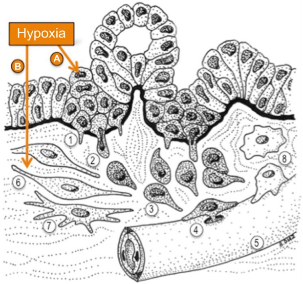
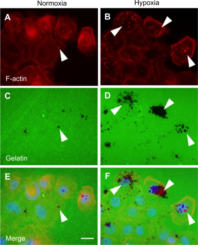
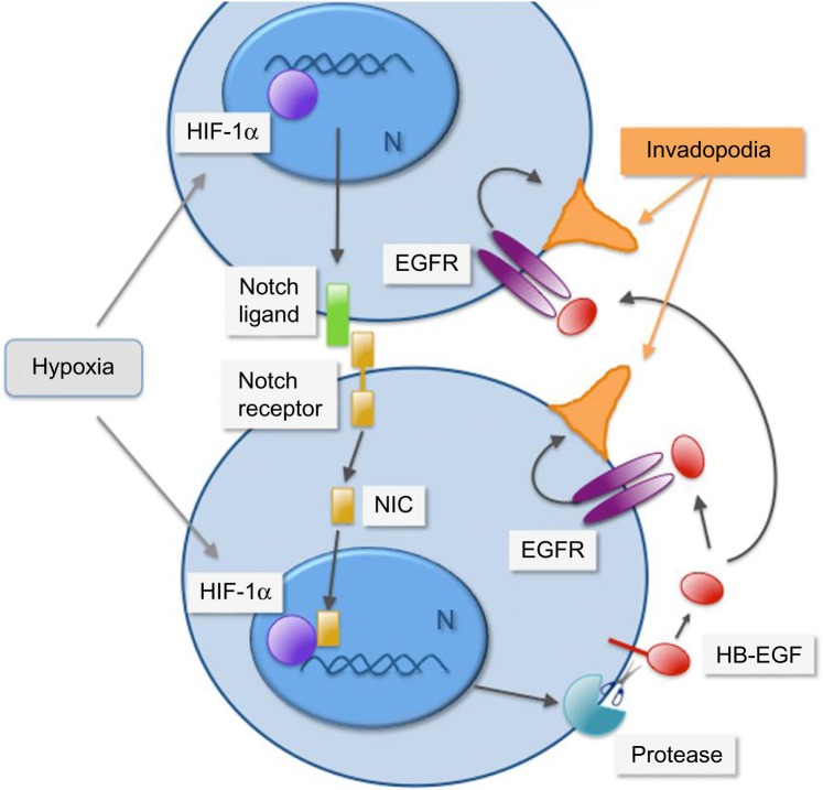
Intratumoral hypoxia is a common feature of solid tumors. Recent advances in cancer biology indicate that hypoxia is not only a consequence of unrestrained tumor growth, but also plays an active role in promoting tumor progression, malignancy, and resistance to therapy. Hypoxia signaling is mediated by the hypoxia-inducible factors (HIFs), which are not only stabilized under hypoxia, but also by activated oncogenes or inactivated tumor suppressors under normoxia. Hypoxia is a prominent feature of the tumor microenvironment of pancreatic tumors, also characterized by the presence of a fibrotic reaction that promotes, and is also modulated by, hypoxia. As the mechanisms by which hypoxia signaling impacts invasion and metastasis in pancreatic cancer are being elucidated, hypoxia is emerging as a key determinant of pancreatic cancer malignancy as well as an important target for therapy. Herein we present an overview of recent advances in the understanding of the impact that hypoxia has in pancreatic cancer invasion and metastasis.

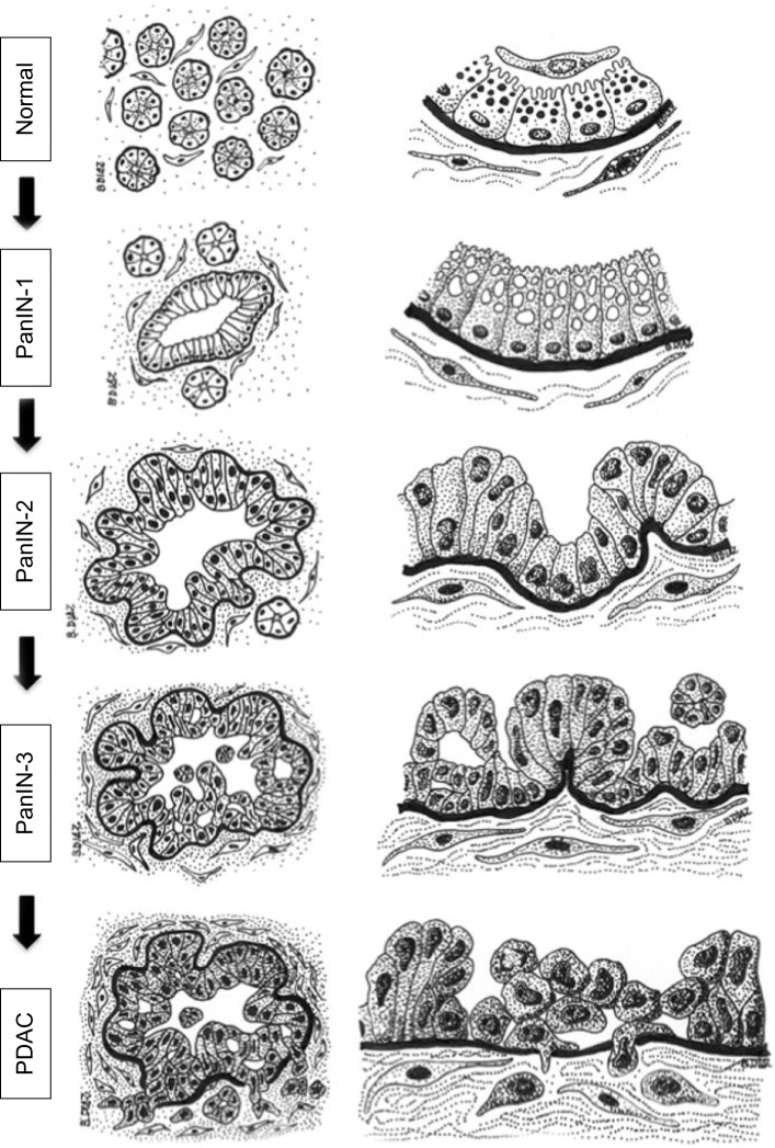
Keywords: EMT; HIF-1; PDAC; PanIN; invadopodia; invasion.


Figures

Figure 1
Figure 1
Diagram of the main anatomic features of the exocrine pancreas. Notes: The exocrine pancreas, which secretes pancreatic juice to the duodenum, is a glandular structure formed by a number of functional units or acini (1). The secretion of each acinus drains into small intercalated ducts (2), which then drain into interlobular ducts (3). Each acinus is formed by the secretory acinar cells (4), which have basally located nuclei and contain abundant zymogen granules in the cytoplasm, whose content is secreted to the center of the acinus through their apical membranes. Acini also contain ductal cells (5) and centroacinar cells in contact with the acinar lumen. Each acinus is surrounded by a basement membrane (clear dotted sheet) that separates the epithelial cells from the stromal component of the gland. The stroma is composed of interstitial fibroblasts (6), pancreatic stellate cells (7) – characterized by the presence of cytoplasmic vitamin A-containing droplets – and a loose interstitial extracellular matrix (8). Sparse blood vessels, also surrounded by a basement membrane, can be found within the interstitial stroma (9) of the normal gland.
Figure 2
Figure 2
Diagram of the histology of precursor lesions and PDAC. Notes: The progression model from normal exocrine pancreas to PDAC is depicted as diagrams of representative tissue sections (left column) and their corresponding magnifications (right column). The normal exocrine pancreas is formed by pancreatic acini surrounded by stromal tissue containing fibroblasts and pancreatic stellate cells. Each acinus contains ductal (not depicted), centroacinar, and acinar cells that contain zymogen granules and are separated from the stroma by a basement membrane (black). Recent studies indicate that acinar cells may be the epithelial cell type targeted by oncogenic transformation during PDAC initiation, although a centroacinar or ductal origin for PDAC cannot be completely ruled out (see text for details). PanIN lesions already contain mutations in K-RAS and are characterized by a columnar epithelium resembling the ductal epithelium. Cells in PanIN-1 often contain mucin in the cytoplasm and present normal nuclei located basally. PanIN-2 lesions are characterized by hyperplasia (excessive cell proliferation), which leads to papillary protrusions into the lumen. The epithelium often becomes pseudostratified, and not all nuclei are basally located, indicating initial loss of epithelial polarity. Nuclei are abnormally shaped. PanIN-3 is the most advanced precursor lesion and displays extensive hyperplasia with papillary protrusions into the lumen, loss of epithelial polarity, and abnormally shaped nuclei that pile up. In PDAC, cells become mesenchymal and invasive, cross the basement membrane, and migrate through the stroma. The integrity of the basement membrane is the main histological feature that differentiates PanIN-3 and PDAC, although recent studies indicate that cells may cross the basement membrane earlier during the progression of the disease (see text for details). Note that the stroma surrounding the lesions and the cancer cells also evolves during the progression of PDAC, becoming more dense and fibrotic. Fibrosis starts in PanIN-1 lesions and progressively increases through PanIN-2 and -3 until the formation of the intense desmoplastic reaction that characterizes PDAC (see text for details). Abbreviations: PanIN, pancreatic intraepithelial neoplasia; PDAC, pancreatic ductal adenocarcinoma.
Figure 3
Figure 3
Hypoxia potentiates invasive steps in PDAC. Notes: Hypoxia in the tumor microenvironment potentiates PDAC cell invasiveness by inducing molecular pathways that affect (A) the cancer cells and (B) the tumor stroma. (A) Hypoxia promotes EMT and also facilitates the invasive migration of cancer cells through extracellular barriers. Upon EMT, epithelial cells lose the attachment to their neighboring cells within the epithelial layer and acquire a migratory phenotype. The process of invasion starts when PDAC cells breach the epithelial basement membrane that separates the epithelium from the stroma (black). Invasion is aided in vitro and possibly in vivo by the formation of proteolytic protrusions named invadopodia (1) that locally digest the basement membrane, generating pores that cancer cells use to cross it and reach the stroma (2). Once in the stroma, PDAC cells display invasive directed migration, aided by EMT and the proteolytic activity of invadopodia (3) toward blood (5) or lymphatic vessels to intravasate and disseminate through the circulation. PDAC cells are likely to use invadopodia to cross endothelial basement membranes (4) and reach the circulation. (B) Hypoxia affects the tumor stroma by promoting the profibrotic activity of cancer-associated fibroblasts (6) and pancreatic stellate cells (7), which further increases hypoxia in the tumor microenvironment, and also by promoting the crosstalk between cancer and stromal cells to increase cancer cells invasiveness (see text for details). Hypoxia may also affect the recruitment and activation of macrophages (8), which also interact with cancer cells to facilitate invasion, for instance, during intravasation. Hypoxia is a determinant of PDAC invasiveness through the activation of cell autonomous and non-cell-autonomous mechanisms that operate in both cancer and associated stroma. Abbreviations: EMT, epithelial-to-mesenchymal transition; PDAC, pancreatic ductal adenocarcinoma.
Figure 4
Figure 4
Hypoxia increases invadopodia formation in human PDAC cells. Notes: The human PDAC line BxPC3 was cultured under normoxia or hypoxia (1% oxygen) for 16 hours on coverslips covered by fluorescein isothiocyanate-conjugated gelatin (green). Cells were processed for detection of invadopodia by F-actin labeling with Alexa468-conjugated phalloidin (red). Nuclei were stained with DAPI (blue). Invadopodia are detected as F-actin-rich dots (arrowheads in A and B) that display local protease activity by digesting the gelatin (black areas indicate absence of gelatin upon invadopodia activity; arrowheads in C and D). Gelatin degradation often co-localizes with invadopodia (arrowheads in E and F). Size bar: 10 µm. Abbreviations: DAPI, 4′,6-diamidino-2-phenylindole; PDAC, pancreatic ductal adenocarcinoma.
Figure 5
Figure 5
Cell-contact-dependent signaling and paracrine activation of the EGF receptor are coupled under hypoxia to promote invadopodia formation in pancreatic cancer cells. Notes: In pancreatic cancer cells, hypoxia promotes HIF-1α-dependent activation of the Notch signaling pathway, which mediates cell-contact-dependent signaling upon binding of a transmembrane ligand to a transmembrane receptor expressed in adjacent cells. Upon ligand binding, the receptor is cleaved by proteolysis to release a Notch intracellular domain (NIC) that travels to the nucleus (N) and partners with HIF-1α to promote gene transcription. Activation of Notch signaling under hypoxia promotes protease-dependent shedding of the EGF receptor ligand HB-EGF (red). Upon shedding, the transmembrane HB-EGF becomes soluble and signals in autocrine, juxtacrine, or paracrine fashions. Activation of the EGF receptor by soluble HB-EGF promotes invadopodia (depicted in orange) formation and increases cancer cell invasiveness. Adapted with permission from Díaz B, Yuen A, Iizuka S, Higashiyama S, Courtneidge SA. Notch increases the shedding of HB-EGF by ADAM12 to potentiate invadopodia formation in hypoxia. J Cell Biol. 2013;201(2):279–292. Abbreviations: EGF, epidermal growth factor; EGFR, epidermal growth factor receptor; HB-EGF, heparin-binding epidermal growth factor; HIF-1α, hypoxia-inducible factor 1α.

Similar articles

Cited by

References

    1. Siegel R, Ward E, Brawley O, Jemal A. Cancer Statistics, 2011: the impact of eliminating socioeconomic and racial disparities on premature cancer deaths. CA Cancer J Clin. 2011;61:212–236. - PubMed
    1. Hidalgo M. Pancreatic cancer. N Engl J Med. 2010;362(17):1605–1617. - PubMed
    1. Vincent A, Herman J, Schulick R, Hruban RH, Goggins M. Pancreatic cancer. Lancet. 2011;378(9791):607–620. - PMC - PubMed
    1. Feig C, Gopinathan A, Neesse A, Chan DS, Cook N, Tuveson DA. The pancreas cancer microenvironment. Clin Cancer Res. 2012;18(16):4266–4276. - PMC - PubMed
    1. Pérez-Mancera PA, Guerra C, Barbacid M, Tuveson DA. What we have learned about pancreatic cancer from mouse models. Gastroenterology. 2012;142(5):1079–1092. - PubMed