. 2017 Jun;127(6):E193-E200.
doi: 10.1002/lary.26333. Epub 2016 Oct 24.

Cellular source and proinflammatory roles of high-mobility group box 1 in surgically injured rat vocal folds

Affiliations

Cellular source and proinflammatory roles of high-mobility group box 1 in surgically injured rat vocal folds

Nicole Y K Li-Jessen et al. Laryngoscope. 2017 Jun.

Abstract

Objectives/hypothesis: High-mobility group box 1 (HMGB1) is a chromatin-binding protein located in the cell nucleus. Following injury, immunocompetent cells secrete HMGB1 to the extracellular milieu under the stimulation of proinflammatory cytokines. Extracellular HMGB1 acts a danger signal that instigates the innate immunity and tissue repair. We previously reported HMGB1 in the vocal fold extracellular compartment between day 3 and day 7 following surgical injury. In this study, we further investigated the cell source of HMGB1 and the relationship of proinflammatory cytokine expression and HMGB1 translocation in wounded vocal folds.

Study design: Prospective animal study.

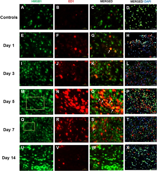
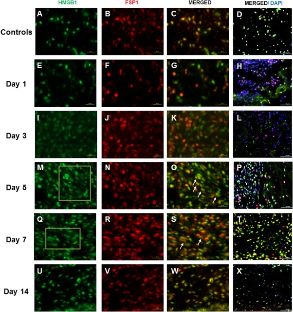
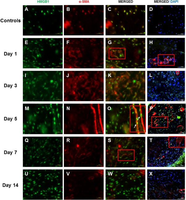
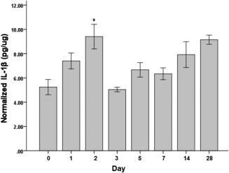
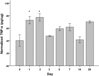
Methods: Bilateral vocal fold injury was performed on 122 Sprague-Dawley rats. An additional 18 rats served as uninjured controls. Animals were sacrificed at multiple time points up to 4 weeks after surgery. Immunohistochemical costaining was performed to identify the cell source of HMGB1. Cell markers ED1, fibroblast-specific protein 1 (FSP1), and alpha smooth muscle actin (α-SMA) were used to identify macrophages, fibroblasts, and myofibroblasts, respectively. Enzyme-linked immunosorbent assays were performed to measure cytokine levels of interleukin-1beta (IL-1β) and tumor necrosis factor-alpha (TNF-α) in vocal fold tissue.

Results: Costaining of HMGB1 was strong with ED1 and FSP1 but was minimal with α-SMA in injured vocal folds. Compared to uninjured controls, IL-1β and TNF-α expression increased significantly the first 2 days after injury.

Conclusions: Macrophages and fibroblasts were a major cell source of vocal fold HMGB1. Translocation of HMGB1 may be an active response to the early accumulation of IL-1β and TNF-α in the wounded vocal folds.

Level of evidence: NA Laryngoscope, 127:E193-E200, 2017.

Keywords: HMGB1; cytokines; inflammation; vocal folds; wound healing.


Figures

Figure 1
Figure 1
High‐mobility group chromosomal box (HMGB1) and ED1 expression following vocal fold surgical injury. (A, E, I, M, Q, U) Representative immunohistochemical (IHC) staining of HMGB1 in uninjured and injured rat vocal folds over 2 weeks postsurgery. HMGB1 is stained green. (B, F, J, N, R, V) Representative IHC staining of ED1 of the same animals. ED1 is stained red. (C, G, K, O, S, W) Representative IHC staining of merged HMGB1 and ED1 of the same animals. (D, H, L, P, T, X) Representative IHC staining of merged HMGB1, ED1, and 4,6‐diamidino‐2‐phenylindole (DAPI) of the same animals. Yellow cells are costained with HMGB1 (green) and ED1 (red). Examples are indicated with white arrows. In addition to nuclear expression, extracellular HMGB1 staining was evident in injured samples at day 5 and day 7 postsurgery. Examples are indicated in green boxes. At day 14, extracellular HMGB1 deposition became less evident. All images were taken at ×630 original magnification (scale bar = 20 μm), except D, H, L, P, T, and X, which were taken at ×400 original magnification (scale bar = 50 μm) to indicate the relative distribution of ED1‐positive cells to the overall cell population.
Figure 2
Figure 2
High‐mobility group chromosomal box (HMGB1) and fibroblast‐specific protein 1 (FSP1) expression following vocal fold surgical injury. (A, E, I, M, Q, U) Representative immunohistochemical (IHC) staining of HMGB1 in uninjured and injured rat vocal folds over 2 weeks postsurgery. HMGB1 is stained green. (B, F, J, N, R, V) Representative IHC staining of FSP1 of the same animals. FSP1 is stained red. (C, G, K, O, S, W) Representative IHC staining of merged HMGB1 and FSP1 of the same animals. (D, H, L, P, T, X) Representative IHC staining of merged HMGB1, FSP1, and 4,6‐diamidino‐2‐phenylindole (DAPI) of the same animals. Yellow cells are costained with HMGB1 (green) and FSP1 (red). Examples are indicated with white arrows. In addition to nuclear expression, extracellular HMGB1 staining was evident in injured samples at day 5 and day 7 postsurgery. Examples are indicated in green boxes. At day 14, extracellular HMGB1 deposition became less evident. All images were taken at ×630 original magnification (scale bar = 20 μm), except D, H, L, P, T, and X, which were taken at ×400 magnification (scale bar = 50 μm) to indicate the relative distribution of FSP1‐positive cells to the overall cell population.
Figure 3
Figure 3
High‐mobility group chromosomal box (HMGB1) and alpha smooth muscle actin (α‐SMA) expression following vocal fold surgical injury. (A, E, I, M, Q, U) Representative immunohistochemical (IHC) staining of HMGB1 of the uninjured and injured rat vocal folds over 2 weeks postsurgery. HMGB1 is stained green. (B, F, J, N, R, V) Representative IHC staining of α‐SMA in the same animals. α‐SMA is stained red. Most α‐SMA–positive cells were found around the blood vessel walls. (C, G, K, O, S, W) Representative IHC staining of merged HMGB1 and α‐SMA of the same animals. (D, H, L, P, T, X) Representative IHC staining of merged HMGB1, α‐SMA, and 4,6‐diamidino‐2‐phenylindole (DAPI) of the same animals. Yellow cells are costained with HMGB1 (green) and α‐SMA (red). Examples are indicated with white arrows. The blood vessel walls are indicated with red boxes. All images were taken at ×630 magnification (scale bar = 20 μm), except D, H, L, P, T, and X, which were taken at ×400 magnification (scale bar = 50 μm) to indicate the relative distribution of α‐SMA‐positive cells to the overall cell population.
Figure 4
Figure 4
Interleukin (IL)‐1β protein levels normalized to the total protein level in the tissue samples following vocal fold surgical injury. IL‐1β concentrations are in picograms per microgram. Bars and error bars represent mean and standard error of the data (n = 32), respectively. Asterisk denotes that data for that time point are statistically significant compared to uninjured controls (Bonferroni‐corrected alpha: .05/7 = .007).
Figure 5
Figure 5
Tumor necrosis factor (TNF)‐α protein levels normalized to the total protein level in the tissue samples following vocal fold surgical injury. TNF‐α concentrations are in picograms per microgram. Bars and error bars represent mean and standard error of the data (n = 32), respectively. Asterisks denote that data for that time point are statistically significant compared to the uninjured controls (Bonferroni‐corrected alpha: .05/7 = .007).

References

    1. Klune JR, Dhupar R, Cardinal J, Billiar TR, Tsung A. HMGB1: endogenous danger signaling. Mol Med 2008;14:476–484. - PMC - PubMed
    1. Foell D, Wittkowski H, Roth J. Mechanisms of disease: a ‘DAMP’ view of inflammatory arthritis. Nat Clin Pract Rheumatol 2007;3:382–390. - PubMed
    1. Wang H, Yang H, Tracey KJ. Extracellular role of HMGB1 in inflammation and sepsis. J Intern Med 2004;255:320–331. - PubMed
    1. Yang H, Tracey KJ. High mobility group box 1 (HMGB1). Crit Care Med 2005;33:S472–S474. - PubMed
    1. Peltz ED, Moore EE, Eckels PC, et al. HMGB1 is markedly elevated within 6 hours of mechanical trauma in humans. Shock 2009;32:17–22. - PMC - PubMed

Publication types

MeSH terms

LinkOut - more resources