

Distribution, physiology and pharmacology of relaxin-3/RXFP3 systems in brain
- PMID: 27774604
- PMCID: PMC5406293
- DOI: 10.1111/bph.13659
Distribution, physiology and pharmacology of relaxin-3/RXFP3 systems in brain
Abstract
Relaxin-3 is a member of a superfamily of structurally-related peptides that includes relaxin and insulin-like peptide hormones. Soon after the discovery of the relaxin-3 gene, relaxin-3 was identified as an abundant neuropeptide in brain with a distinctive topographical distribution within a small number of GABAergic neuron populations that is well conserved across species. Relaxin-3 is thought to exert its biological actions through a single class-A GPCR - relaxin-family peptide receptor 3 (RXFP3). Class-A comprises GPCRs for relaxin-3 and insulin-like peptide-5 and other peptides such as orexin and the monoamine transmitters. The RXFP3 receptor is selectively activated by relaxin-3, whereas insulin-like peptide-5 is the cognate ligand for the related RXFP4 receptor. Anatomical and pharmacological evidence obtained over the last decade supports a function of relaxin-3/RXFP3 systems in modulating responses to stress, anxiety-related and motivated behaviours, circadian rhythms, and learning and memory. Electrophysiological studies have identified the ability of RXFP3 agonists to directly hyperpolarise thalamic neurons in vitro, but there are no reports of direct cell signalling effects in vivo. This article provides an overview of earlier studies and highlights more recent research that implicates relaxin-3/RXFP3 neural network signalling in the integration of arousal, motivation, emotion and related cognition, and that has begun to identify the associated neural substrates and mechanisms. Future research directions to better elucidate the connectivity and function of different relaxin-3 neuron populations and their RXFP3-positive target neurons in major experimental species and humans are also identified.
Linked articles: This article is part of a themed section on Recent Progress in the Understanding of Relaxin Family Peptides and their Receptors. To view the other articles in this section visit http://onlinelibrary.wiley.com/doi/10.1111/bph.v174.10/issuetoc.
© 2016 The British Pharmacological Society.
Figures
 ) or putative untested (
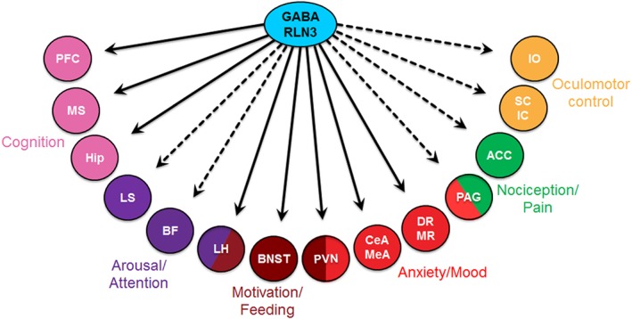
 ) functional roles of relaxin‐3/RXFP3 signalling in the coordinated regulation of modalities including cognition, arousal, motivation, anxiety, mood, pain and oculomotor control. Abbreviations: ACC, anterior cingulate cortex; BF, basal forebrain; CeA, central amygdala; DR, dorsal raphe; Hip, hippocampus; IC, inferior colliculus; IO, inferior olive; LH, lateral hypothalamus; LS, lateral septum; MeA, medial amygdala; MR, median raphe; MS, medial septum; PAG, periaqueductal grey; PFC, prefrontal cortex; PVN, paraventricular hypothalamic nucleus; SC, superior colliculus.
References
-
- Albert‐Gasco H, Garcia‐Aviles A, Moustafa S, Sanchez‐Sarasua S, Gundlach AL, Olucha‐Bordonau FE et al. (2016). Central relaxin‐3 receptor (RXFP3) activation increases ERK phosphorylation in septal cholinergic neurons and impairs spatial working memory. Brain Struct Funct. doi:10.1007/s00429-016-1227-8. - DOI - PubMed
-
- Banerjee A, Shen PJ, Ma S, Bathgate RAD, Gundlach AL (2010). Swim stress excitation of nucleus incertus and rapid induction of relaxin‐3 expression via CRF1 activation. Neuropharmacology 58: 145–155. - PubMed
-
- Bathgate RAD, Ivell R, Sanborn BM, Sherwood OD, Summers RJ (2006a). International union of pharmacology LVII: recommendations for the nomenclature of receptors for relaxin family peptides. Pharmacol Rev 58: 7–31. - PubMed
Publication types
MeSH terms
Substances
LinkOut - more resources
Full Text Sources
Other Literature Sources
Molecular Biology Databases
