Review
. 2016 Dec;38(12):1234-1245.
doi: 10.1002/bies.201600154. Epub 2016 Oct 24.

Centriole positioning in epithelial cells and its intimate relationship with planar cell polarity

Affiliations
Review

Centriole positioning in epithelial cells and its intimate relationship with planar cell polarity

Jose Maria Carvajal-Gonzalez et al. Bioessays. 2016 Dec.

Abstract

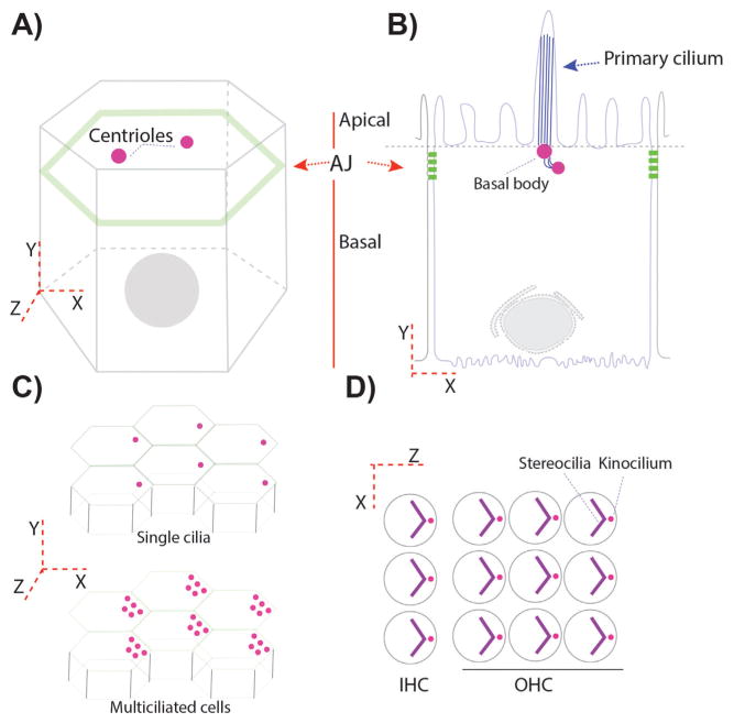
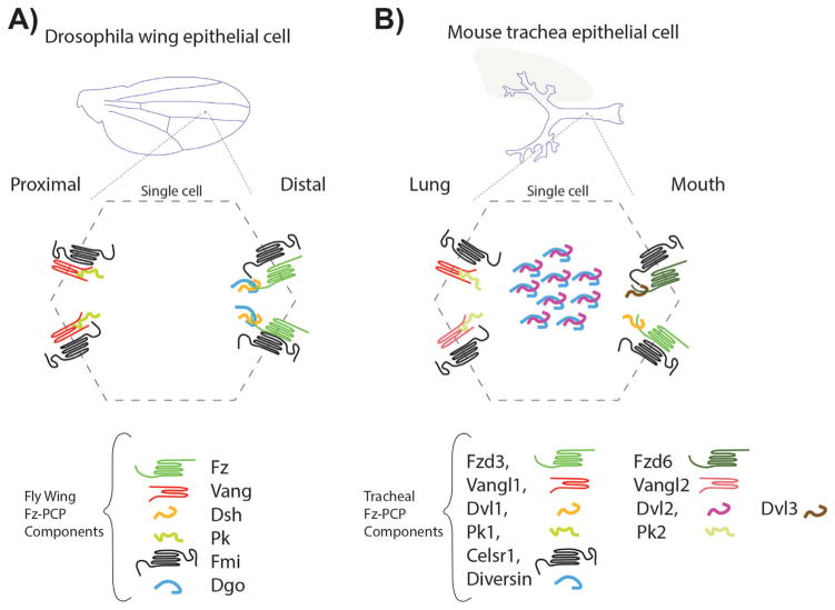
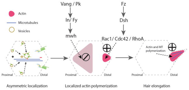
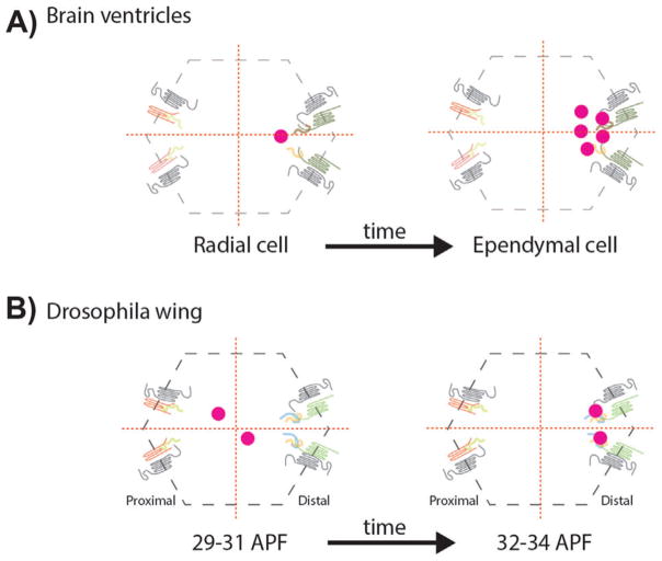
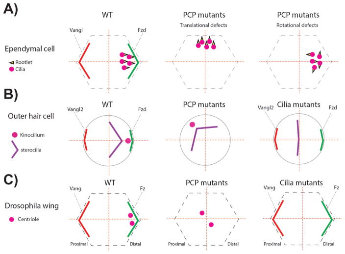
Planar cell polarity (PCP)-signaling and associated tissue polarization are evolutionarily conserved. A well documented feature of PCP-signaling in vertebrates is its link to centriole/cilia positioning, although the relationship of PCP and ciliogenesis is still debated. A recent report in Drosophila established that Frizzled (Fz)-PCP core signaling has an instructive input to polarized centriole positioning in non-ciliated Drosophila wing epithelia as a PCP read-out. Here, we review the impact of this observation in the context of recent descriptions of the relationship(s) of core Fz-PCP signaling and cilia/centriole positioning in epithelial and non-epithelial cells. All existing data are consistent with a model where Fz-PCP signaling functions upstream of centriole/cilia positioning, independent of ciliogenesis. The combined data sets indicate that the Fz-Dsh PCP complex is instructive for centriole/ciliary positioning via an actin-based mechanism. Thereby, centriole/cilia/centrosome positioning can be considered an evolutionarily conserved readout and common downstream effect of PCP-signaling from flies to mammals.

Keywords: Frizzled-Vangl signaling; actin cytoskeleton; centriole/MTOC localization; cilia; epithelial polarity; microtubules; planar cell polarity.


Figures

Figure 1
Figure 1
Centriole/basal body positioning in polarized epithelial cells. A and B: Epithelial cells are like prisms with a belt of proteins connecting adjacent cells. These proteins form adherens junctions (AJ) in Drosophila (A) and AJ and tight junctions (TJ) in vertebrates (B). AJ or AJ/TJ are responsible for delimiting the apical and basal membrane domains in polarized epithelial cells. Centrioles, in polarized epithelial cells, are localized in the most apical planes (A and B). B: In a ciliated epithelium, centrioles migrate towards the apical membrane, where they contact the apical membrane becoming the “basal bodies”/BB, from which the cilia form and protrude into the apical space. C: Certain epithelia display a tissue-coordinated off-center positioning of BBs/centrioles, but still within the most apical planes. This is also observed in many multiciliated epithelia (bottom graph). D: In more specialized epithelial cells, like the outer and inner sensory hair cells (OHC and IHC) in the organ of Corti, the primary cilium, called kinocilium here, is also polarized in an off-center position in the apical plane, subsequently organizing an actin based V-shaped structure, the stereocilia bundle, which follows the polarization of the kinocilium.
Figure 2
Figure 2
Frizzled (Fz) planar cell polarity core components in Drosophila wings and mouse trachea. A: Drosophila wing blades are formed by two layers of polarized epithelial cells with the apical domain facing the wing surface. Following the proximal-distal axis, Fz-PCP core components are asymmetrically localized to a distal complex including Fz-Dsh-Fmi-Dgo and a proximal complex formed by Vang-Pk-Fmi. This pattern is reproduced in every single cell throughout the wing blade. B: In mouse tracheal epithelial cells, the apical domains face the lumen. In an analogous manner to Drosophila wing epithelial cells, the Fz-PCP core components are segregated in proximal (lung side) and distal (mouth side) complexes, containing the equivalent orthologues as in Drosophila. In addition to the several isoforms for most of the Fz-PCP core components, in mouse tracheal epithelial cells Diversin (the vertebrate Dgo homolog) and Dvl2 are detected near the basal bodies in these multiciliated cells.
Figure 3
Figure 3
Cytoskeletal arrangements during the planar cell polarization process in Drosophila pupal wing epithelial cells. uring pupal development, epithelial cells use essential cellular machinery, including the protein trafficking machinery which is involved in the asymmetric localization of the Fz-PCP core components in a polarized manner. This directed transport requires an alignment of well oriented microtubules (MT, blue line in left panel) with enriched minus-ends towards the distal side of each cell (left panel). This stage is followed by activation of actin polymerization in the distal side (via Fz-Dsh signaling), while blocking actin polymerization in the proximal side (via Vang-Pk and their effectors) of the same cell (middle panel). The last stage involves actin and stable microtubule polymerization at the base and inside trichomes (cellular hairs), which is essential for hair elongation (right panel).
Figure 4
Figure 4
Planar polarized centriole positioning. A: Centrioles, as basal bodies (BBs) at the apical plane lining the brain ventricle cells, are planar cell polarized, first in radial cells and followed by their differentiation to ependymal cells, where BBs are multiplied generating a multiciliated epithelium. The off-centered distribution in both, radial and ependymal cells, is controlled by the Fz-PCP signaling pathway. B: During Drosophila pupal wing development, centrioles get polarized along the Fz-PCP polarization axis, migrating towards the Fz-Dsh-Fmi-Dgo distal domain. APF: After puparium formation. Protein cartoons are as in Fig. 2 with the Fz-Dsh/Dvl complexes on the right in each cell.
Figure 5
Figure 5
PCP and cilia mutants associated phenotypes related to centriole/basal body (BB) A: In ependymal cells BBs are localized near the Fzd expression domain. In addition, the basal foot of the cilia, called rootlet and critical for ciliary beating, is aligned in the same direction (left panel). In one set of specific PCP mutants (check text for specific mutants) the localization of the BBs leading to so-called “translational defects” (middle panel). In other PCP mutants, although the translational polarity is correct, the alignment of the ciliary rootlets is uncoordinated (right panel). B: In the inner ear, outer hair cells (OHCs) position the BBs of the kinocilium (a specialized primary cilium) near the Fzd-Dvl localization domain, similar to ependymal cells (left panel). In PCP mutants the off-centered positioning of the BB is affected, but the V-shaped stereocilia bundle is not affected, following the position of the BB/kinocilium (middle panel). In cilia mutants, the V-shape of the stereocilia is affected and the structure remains central. The stereocilia bundle can appear as a line or circular shape depending on the severity of the ciliary mutant, in which the kinoclium is missing (right panel). C: Once fully polarized in Drosophila pupal wing cells, centrioles are localized near the Fz-Dsh domain, similar to mouse ependymal cells and OHCs (left panel). In PCP core and effector mutants, centrioles remain centered (middle panel). In Sas4 mutants where centrioles are absent, PCP remains mostly normal and actin polymerization in trichomes is largely unaffected (right panel).

Similar articles

Cited by

References

    1. Bornens M. The centrosome in cells and organisms. Science. 2012;335:422–6. - PubMed
    1. Carvalho-Santos Z, Azimzadeh J, Pereira-Leal JB, Bettencourt-Dias M. Evolution: tracing the origins of centrioles, cilia, and flagella. J Cell Biol. 2011;194:165–75. - PMC - PubMed
    1. Azimzadeh J, Marshall WF. Building the centriole. Curr Biol. 2010;20:R816–25. - PMC - PubMed
    1. Bettencourt-Dias M, Glover DM. Centrosome biogenesis and function: centrosomics brings new understanding. Nat Rev Mol Cell Biol. 2007;8:451–63. - PubMed
    1. Beisson J, Wright M. Basal body/centriole assembly and continuity. Curr Opin Cell Biol. 2003;15:96–104. - PubMed

Publication types

Substances

LinkOut - more resources