. 2017 Feb;26(2):365-374.
doi: 10.1002/pro.3073. Epub 2016 Nov 2.

The macro domain as fusion tag for carrier-driven crystallization

Affiliations

The macro domain as fusion tag for carrier-driven crystallization

Rebekka Wild et al. Protein Sci. 2017 Feb.

Abstract

Obtaining well-ordered crystals remains a significant challenge in protein X-ray crystallography. Carrier-driven crystallization can facilitate crystal formation and structure solution of difficult target proteins. We obtained crystals of the small and highly flexible SPX domain from the yeast vacuolar transporter chaperone 4 (Vtc4) when fused to a C-terminal, non-cleavable macro tag derived from human histone macroH2A1.1. Initial crystals diffracted to 3.3 Å resolution. Reductive protein methylation of the fusion protein yielded a new crystal form diffracting to 2.1 Å. The structures were solved by molecular replacement, using isolated macro domain structures as search models. Our findings suggest that macro domain tags can be employed in recombinant protein expression in E. coli, and in carrier-driven crystallization.

Keywords: carrier-driven crystallization; crystallization tag; histone macroH2A; macro domain; recombinant protein expression.


Figures

Figure 1
Figure 1
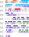
The VTC complex harbors N‐terminal SPX domains. Structure based sequence alignment of VTC subunits ScVtc2 (S. cerevisiae Vtc2; UniProt Acc. P43585), ScVtc3 (S. cerevisiae Vtc3; Q02725) and ScVtc4 (S. cerevisiae Vtc4; P47075) and including a secondary structure assignment calculated with the program DSSP48 and based on PDB entry 5IIG.14 Invariant and conserved residues are highlighted in dark‐ and light‐purple, respectively. SPXScVtc 2 shows 62% and 32% sequence identity with SPXScVtc 3 and SPXScVtc 4, respectively. The two long core helices of the SPX domain are colored in light blue, surrounding helices are colored from yellow to red. The C‐terminal catalytic TTM domain (α‐helices in green, β‐strands in blue) is connected to the SPX domain via a variable linker (in yellow).
Figure 2
Figure 2
A stable SPXScVtc4‐macro fusion protein can be recombinantely expressed in E. coli. (A) Four SPXScVtc 4 constructs were assayed for crystallization, all of which contained a C‐terminal 6xHis tag and either no fusion protein, a macro tag (macro domain of the human histone macroH2A1.126, 27), trxA (E. coli thioredoxinA11) or the catalytic TTM domain22, 23 of ScVtc4. (B) Overview of the constructed macro domain expression plasmid providing a macro tag (human macroH2A1.1 residues 181‐366) connected to the N‐terminal SPX domain via a Ala‐Gly‐Ser linker and followed by a non‐cleavable 6xHis tag. Target proteins can be inserted using the restriction enzymes XbaI/BamHI. (C) SDS‐PAGE analysis of purified SPXScVtc4−6xHis and SPXScVtc4‐macro‐6xHis fusion proteins expressed in E. coli.
Figure 3
Figure 3
The known macro domain structures allow for structure solution of SPXScVtc 4 by molecular replacement. (A) Structural superposition of different, previously described macro H2A1.1 macro domain structures indicates that N‐ and C‐termini are flexible. R.m.s.d. is between 0.7‐2.6 Å comparing 180 corresponding Cα atoms. (B) Table summary of molecular replacement calculations using the different known macroH2A1.1 structures. 1xRFZ: rotation function Z‐score for the 1st molecule to be placed. 1xTFZ: translation function Z‐score for the 1st molecule to be placed. 2xRFZ: rotation function Z‐score for the 2nd molecule to be placed. 2xTFZ: translation function Z‐score for the 2nd molecule. LLG: Log Likelihood Gain of the refined solution. (C) Cα traces of two SPXScVtc4‐macro molecules (SPX domain are shown in light‐blue and yellow, the C‐terminal macro domains in dark‐blue and orange, respectively) related by a pseudo 2‐fold axis in the asymmetric unit of the I212121 crystal form. The central core helices of the SPX domains are highlighted (ribbon diagrams).
Figure 4
Figure 4
Reductively and non‐methylated SPXScVtc4‐macro protein adopts different conformations. (A) Cα trace of the SPXScVtc4‐macro tetramer in the asymmetric unit of the P21 crystal form. The C‐terminal helix of the SPX domain is highlighted in red. The C‐terminal helix of human SPXHsXPR1 has a similar configuration, again contacting a symmetry‐related molecule (PDB‐ID 5IJH14). Comparison of (B) non‐methylated and (C) reductively methylated SPXScVtc4‐macro fusion proteins (in ribbon representation). The zoom‐in shows the connecting Ala‐Gly‐Ser linker in bonds representation (colored in yellow) and including a 2Fo‐Fc omit electron density map contoured at 1.0 σ (blue mesh). (D) Reductively methylated and (E) non‐methylated SPXScVtc4‐macro fusion proteins form different crystal lattices, involving SPX–SPX, macro–macro and SPX–macro interactions. Colors are as in (A) and Figure 3(C).

References

    1. Donahue JP, Patel H, Anderson WF, Hawiger J (1994) Three‐dimensional structure of the platelet integrin recognition segment of the fibrinogen gamma chain obtained by carrier protein‐driven crystallization. Proc Natl Acad Sci USA 91:12178–12182. - PMC - PubMed
    1. Lim K, Ho JX, Keeling K, Gilliland GL, Ji X, Rüker F, Carter DC (1994) Three‐dimensional structure of Schistosoma japonicum glutathione S‐transferase fused with a six‐amino acid conserved neutralizing epitope of gp41 from HIV. Protein Sci 3:2233–2244. - PMC - PubMed
    1. Waugh DS (2016) Crystal structures of MBP fusion proteins. Protein Sci 25:559–571. - PMC - PubMed
    1. Ware S, Donahue JP, Hawiger J, Anderson WF (1999) Structure of the fibrinogen gamma‐chain integrin binding and factor XIIIa cross‐linking sites obtained through carrier protein driven crystallization. Protein Sci 8:2663–2671. - PMC - PubMed
    1. Kliche W, Fujita‐Becker S, Kollmar M, Manstein DJ, Kull FJ (2001) Structure of a genetically engineered molecular motor. EMBO J 20:40–46. - PMC - PubMed

MeSH terms