

Phase Transition of the Bacterium upon Invasion of a Host Cell as a Mechanism of Adaptation: a Mycoplasma gallisepticum Model
- PMID: 27775027
- PMCID: PMC5075909
- DOI: 10.1038/srep35959
Phase Transition of the Bacterium upon Invasion of a Host Cell as a Mechanism of Adaptation: a Mycoplasma gallisepticum Model
Abstract
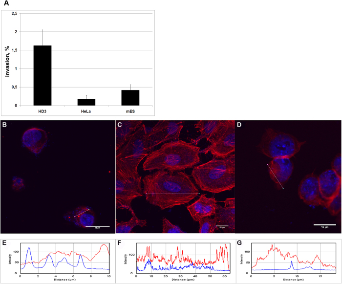
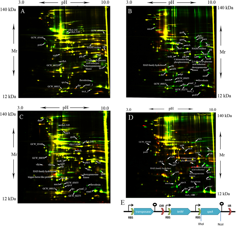
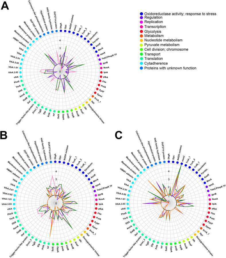
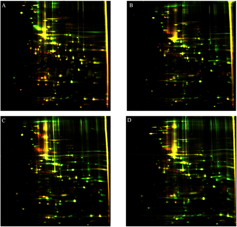
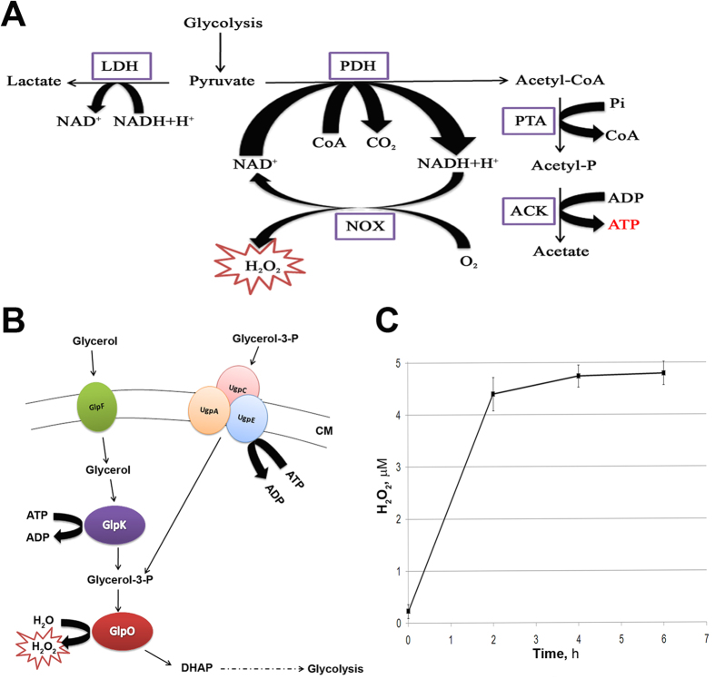
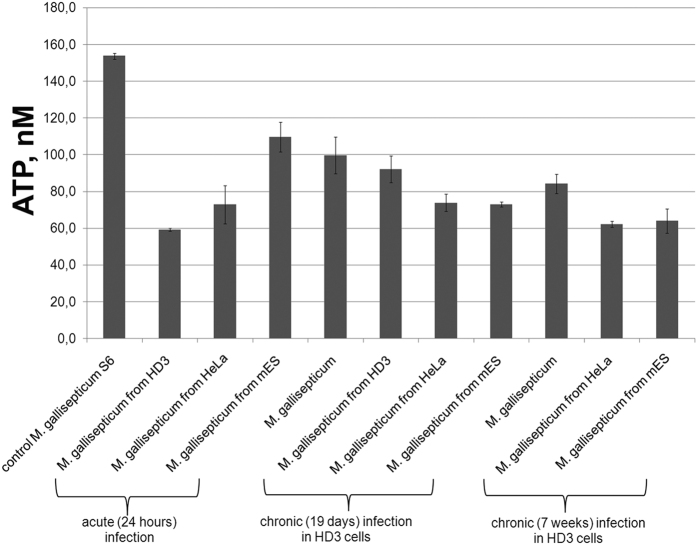
What strategies do bacteria employ for adaptation to their hosts and are these strategies different for varied hosts? To date, many studies on the interaction of the bacterium and its host have been published. However, global changes in the bacterial cell in the process of invasion and persistence, remain poorly understood. In this study, we demonstrated phase transition of the avian pathogen Mycoplasma gallisepticum upon invasion of the various types of eukaryotic cells (human, chicken, and mouse) which was stable during several passages after isolation of intracellular clones and recultivation in a culture medium. It was shown that this phase transition is manifested in changes at the proteomic, genomic and metabolomic levels. Eukaryotic cells induced similar proteome reorganization of M. gallisepticum during infection, despite different origins of the host cell lines. Proteomic changes affected a broad range of processes including metabolism, translation and oxidative stress response. We determined that the activation of glycerol utilization, overproduction of hydrogen peroxide and the upregulation of the SpxA regulatory protein occurred during intracellular infection. We propose SpxA as an important regulator for the adaptation of M. gallisepticum to an intracellular environment.
Figures

Similar articles
-
Transcriptional responses of Mycoplasma gallisepticum strain R in association with eukaryotic cells.J Bacteriol. 2007 Aug;189(16):5803-7. doi: 10.1128/JB.00667-07. Epub 2007 Jun 8. J Bacteriol. 2007. PMID: 17557819 Free PMC article.
-
Expression and immunological characteristics of the surface-localized pyruvate kinase in Mycoplasma gallisepticum.Microb Pathog. 2015 Dec;89:161-8. doi: 10.1016/j.micpath.2015.10.005. Epub 2015 Oct 9. Microb Pathog. 2015. PMID: 26456557
-
Mycoplasma gallisepticum modifies the pathogenesis of influenza A virus in the avian tracheal epithelium.Int J Med Microbiol. 2016 May;306(3):174-86. doi: 10.1016/j.ijmm.2016.04.001. Epub 2016 Apr 6. Int J Med Microbiol. 2016. PMID: 27079856
-
Avian mycoplasmosis (Mycoplasma gallisepticum).Rev Sci Tech. 2000 Aug;19(2):425-42. Rev Sci Tech. 2000. PMID: 10935272 Review.
-
Lag Phase Is a Dynamic, Organized, Adaptive, and Evolvable Period That Prepares Bacteria for Cell Division.J Bacteriol. 2019 Mar 13;201(7):e00697-18. doi: 10.1128/JB.00697-18. Print 2019 Apr 1. J Bacteriol. 2019. PMID: 30642990 Free PMC article. Review.
Cited by
-
Mycoplasma gallisepticum Infection Impaired the Structural Integrity and Immune Function of Bursa of Fabricius in Chicken: Implication of Oxidative Stress and Apoptosis.Front Vet Sci. 2020 Apr 24;7:225. doi: 10.3389/fvets.2020.00225. eCollection 2020. Front Vet Sci. 2020. PMID: 32391391 Free PMC article.
-
A "plus one" strategy impacts replication of felid alphaherpesvirus 1, Mycoplasma and Chlamydia, and the metabolism of coinfected feline cells.mSystems. 2024 Oct 22;9(10):e0085224. doi: 10.1128/msystems.00852-24. Epub 2024 Sep 24. mSystems. 2024. PMID: 39315777 Free PMC article.
-
Proteomic dataset: Profiling of cultivated Echerichia coli isolates from Crohn's disease patients and healthy individuals.Data Brief. 2019 Mar 7;23:103734. doi: 10.1016/j.dib.2019.103734. eCollection 2019 Apr. Data Brief. 2019. PMID: 31372401 Free PMC article.
-
Comparative Proteomic Analysis of Mycoplasma hominis Grown on Media with Different Carbon Sources.Bull Exp Biol Med. 2021 Aug;171(4):449-452. doi: 10.1007/s10517-021-05247-8. Epub 2021 Sep 20. Bull Exp Biol Med. 2021. PMID: 34542749
-
Immune Evasion of Mycoplasma gallisepticum: An Overview.Int J Mol Sci. 2024 Feb 29;25(5):2824. doi: 10.3390/ijms25052824. Int J Mol Sci. 2024. PMID: 38474071 Free PMC article. Review.
References
-
- Naumann M., Schüssler A. & Bonfante P. The obligate endobacteria of arbuscular mycorrhizal fungi are ancient heritable components related to the Mollicutes. ISME J. 4, 862–871 (2010). - PubMed
-
- Werren J. H., Baldo L. & Clark M. E. Wolbachia: master manipulators of invertebrate biology. Nat. Rev Microbiol 6, 741–751 (2008). - PubMed
Publication types
MeSH terms
Substances
LinkOut - more resources
Full Text Sources
Other Literature Sources
