. 2016 Nov 8;55(44):6087-6099.
doi: 10.1021/acs.biochem.6b00986. Epub 2016 Oct 27.

Single-Stranded Nucleic Acids Bind to the Tetramer Interface of SAMHD1 and Prevent Formation of the Catalytic Homotetramer

Affiliations

Single-Stranded Nucleic Acids Bind to the Tetramer Interface of SAMHD1 and Prevent Formation of the Catalytic Homotetramer

Kyle J Seamon et al. Biochemistry. .

Abstract

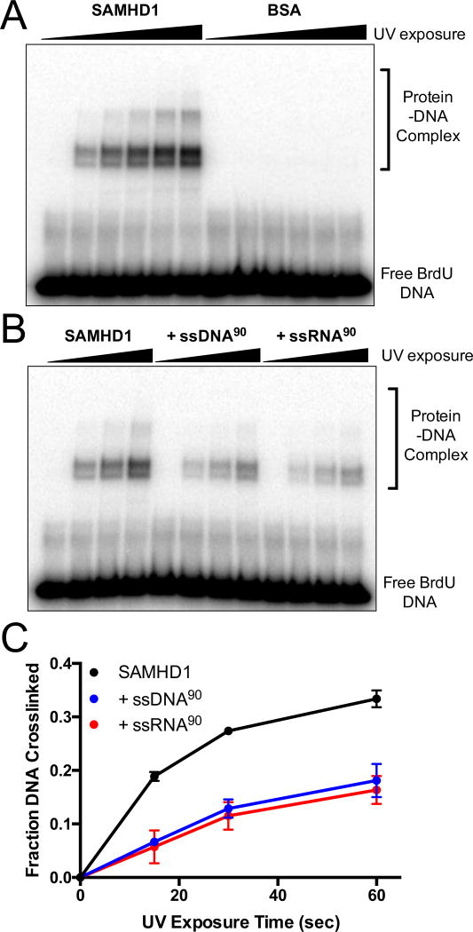
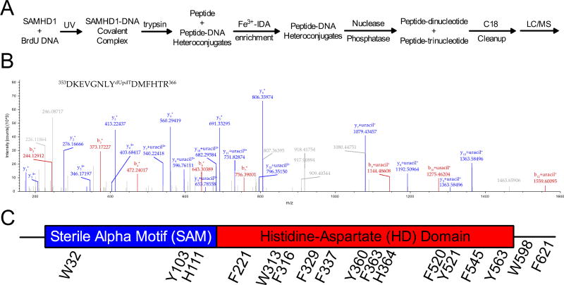
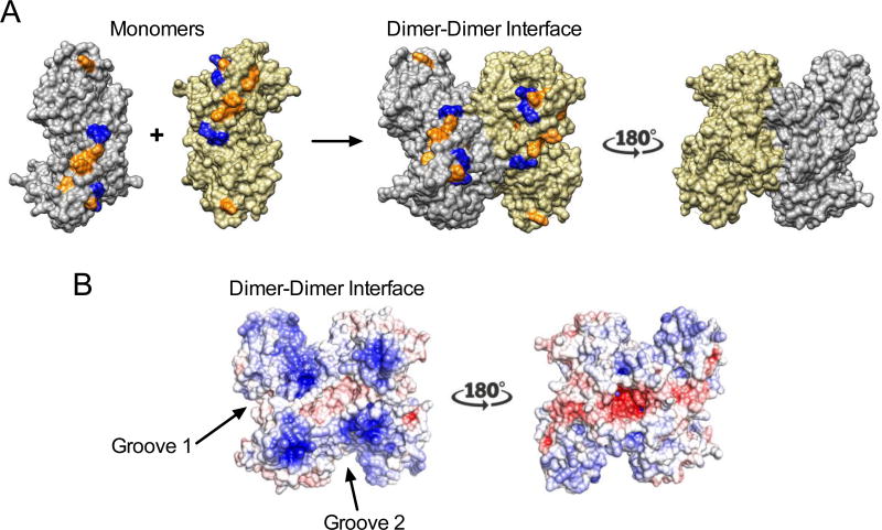
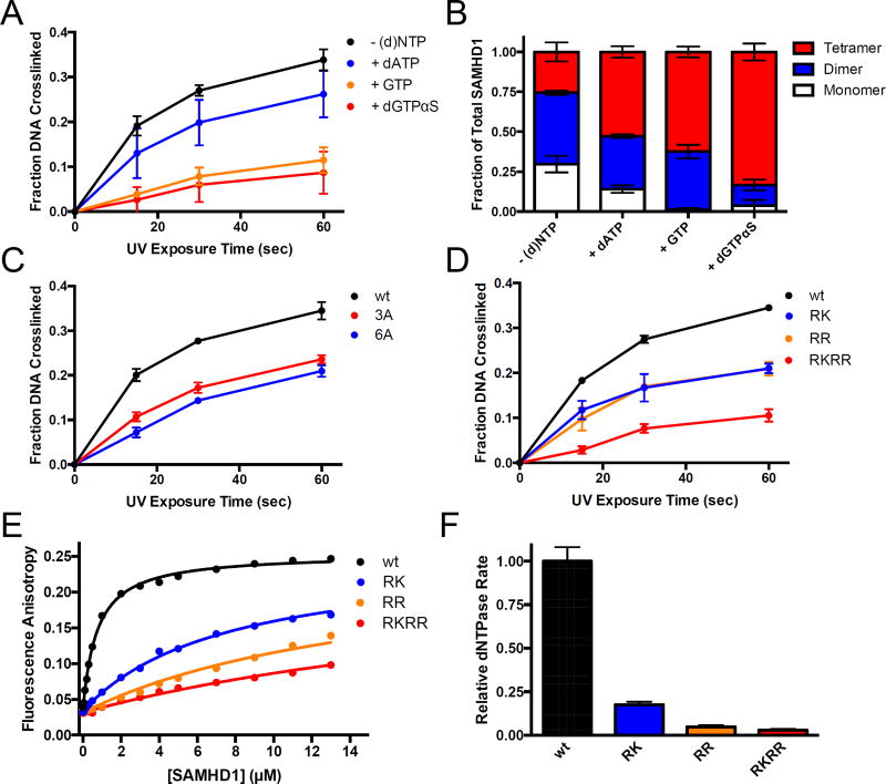
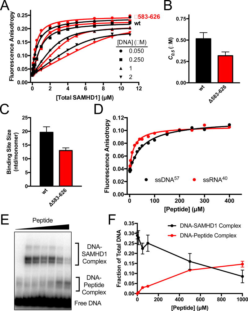
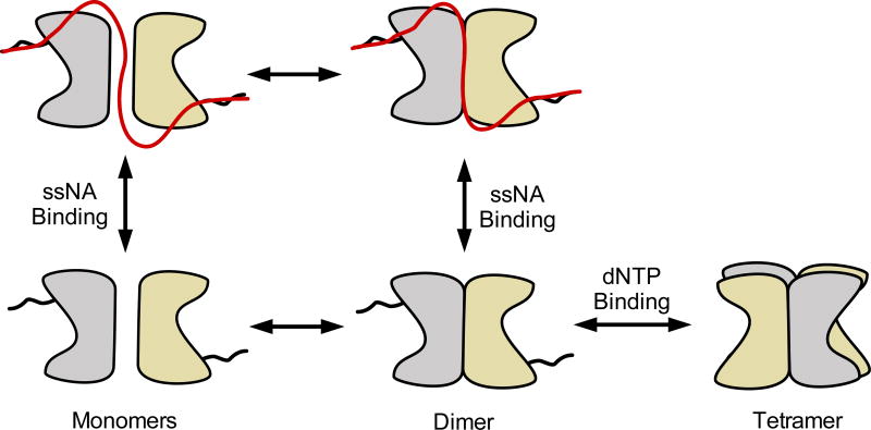
Sterile alpha motif and HD domain protein 1 (SAMHD1) is a unique enzyme that plays important roles in nucleic acid metabolism, viral restriction, and the pathogenesis of autoimmune diseases and cancer. Although much attention has been focused on its dNTP triphosphohydrolase activity in viral restriction and disease, SAMHD1 also binds to single-stranded RNA and DNA. Here we utilize a UV cross-linking method using 5-bromodeoxyuridine-substituted oligonucleotides coupled with high-resolution mass spectrometry to identify the binding site for single-stranded nucleic acids (ssNAs) on SAMHD1. Mapping cross-linked amino acids on the surface of existing crystal structures demonstrated that the ssNA binding site lies largely along the dimer-dimer interface, sterically blocking the formation of the homotetramer required for dNTPase activity. Surprisingly, the disordered C-terminus of SAMHD1 (residues 583-626) was also implicated in ssNA binding. An interaction between this region and ssNA was confirmed in binding studies using the purified SAMHD1 583-626 peptide. Despite a recent report that SAMHD1 possesses polyribonucleotide phosphorylase activity, we did not detect any such activity in the presence of inorganic phosphate, indicating that nucleic acid binding is unrelated to this proposed activity. These data suggest an antagonistic regulatory mechanism in which the mutually exclusive oligomeric state requirements for ssNA binding and dNTP hydrolase activity modulate these two functions of SAMHD1 within the cell.


Figures

Figure 1
Figure 1. Bromodeoxyuridine (BrdU) specifically crosslinks SAMHD1
(A) SAMHD1 or BSA (5 µM) and 32P-labeled BrdU-DNA (5 µM) were mixed, exposed to UV light for 0 to 2 minutes, and analyzed by SDS-PAGE. (B) SAMHD1 (5 µM) and 32P-labeled BrdU DNA (5 µM) were UV irradiated in the presence or absence of unlabeled competitor ssDNA or ssRNA 90mer (5 µM), and analyzed by SDS PAGE. (C) Densitometry of the bands for the free DNA and DNA-SAMHD1 complexes in (B) was used to calculate the fraction of BrdU DNA crosslinked to SAMHD1.
Figure 2
Figure 2. Mass spectrometry for the identification of BrdU crosslinks to SAMHD1
(A) A schematic for mass spectrometry sample preparation. (B) A representative MS2 spectrum from a cross-linked peptide. The parent ion m/z corresponded to the shown peptide with an attached dUpdT dinucleotide and the shown fragment ions unambiguously identify Y360 as the crosslinked residue. Upon fragmentation, b and y ions of the peptide were observed, and the di- or trinucleotide modification fragmented to uracil. (C) All of the cross-linked amino acids observed in the mass spectrometry studies are shown on the linear domain architecture of SAMHD1.
Figure 3
Figure 3. Structural analysis of the BrdU cross-linked regions
(A) Cross-linked aromatic amino acids (orange) are mapped onto the crystal structure of SAMHD1 (PDB 3U1N): F221, W313, F316, F337, Y360, H364, F520, F545, Y563. The Arg residues that have been mutated in this study are shown in blue (R333, K336, R371, R372). (B) Electrostatic surface potential of the HD domain dimer shown using the same two views in (A). The potential was obtained from Poisson-Boltzmann (PB) electrostatics calculations using the APBS tool in Chimera. The two grooves that are flanked by the crosslinking sites in the monomers and dimer are indicated.
Figure 4
Figure 4. Biochemical validation of the mass spectrometry crosslinking results
(A) BrdU crosslinking reactions were performed with SAMHD1 in the presence of dATP, GTP, or dGTPαS to induce oligomerization (1 mM each) and the fraction of total DNA crosslinked to SAMHD1 was quantified. (B) The oligomeric state of SAMHD1 under the conditions in (A) was assessed by glutaraldehyde crosslinking and quantified by densitometry. (C) BrdU crosslinking reactions were performed with wild-type SAMHD1, or the 3A and 6A alanine mutants (see text) and the fraction of the total DNA cross-linked to the protein was quantified. (D) BrdU crosslinking reactions with wild-type SAMHD1 or the indicated glutamate charge-reversal mutants RK, RR, and RKRR (see text). The fraction of the total DNA cross-linked to the protein at each time point was quantified by densitometry. (E) Increasing anisotropy of a fluorescein-labeled dT60 DNA oligonucleotide as a function of added wt or mutant SAMHD1. (F) The normalized dNTP hydrolase activity of wt SAMHD1 and the indicated mutants was determined using 1 mM dGTP as the substrate.
Figure 5
Figure 5. The disordered C-Terminus of SAMHD1 binds ssNA
(A) Fluorescence anisotropy titrations of the fluorescein-labeled dT60 with wt and Δ583–626 SAMHD1 as a function of DNA concentration. The data were fit to a variable stoichiometry quadratic binding equation. (B) Comparisons of the concentrations of wt and Δ583–626 SAMHD1 required for half-maximal DNA binding (C0.5). (C) Comparison of the calculated binding site sizes of wt SAMHD1 and Δ583–626 SAMHD1. The sizes are indicated as nt/monomer. (D) Fluorescence anisotropy increases of FAM-dT60 and FAM-ssRNA upon binding of a peptide consisting of residues 582–626 of SAMHD. (E) BrdU crosslinking reactions were performed with fixed concentrations of BrdU DNA (5 µM) and SAMHD1 (5 µM), and variable concentrations (0 to 1 mM) of the 582–626 SAMHD1 peptide. (F) Quantification of the DNA-SAMHD1 and DNA-peptide complexes from (F) by densitometry.
Figure 6
Figure 6. SAMHD1 does not posses hydrolytic or phosphorolytic RNase Activity
(A) Reactions of 5’ 32P-labeled ssRNA 20mer (1 µM) and SAMHD1 (0.5 µM) were carried out in Tris/KCl/MgCl2 buffer supplemented with the indicated concentration of phosphate and degradation products were resolved by denaturing PAGE. (B) Reactions of 3H-dGTP (1 mM) and SAMHD1 (0.5 µM) carried out under the same conditions as above and the products were resolved by RP-TLC. (C) Reactions were prepared as in (A) but with 0.1 µM human polynucleotide phosphorylase as a positive control. Robust phosphorolytic activity is observed with 10 mM Pi, but is inhibited by high concentrations of Pi as previously reported (D) Reactions prepared with Tris/KCl/MgCl2 buffer, 32Pi (10 mM), poly(A) RNA (1 mg/mL), and SAMHD1 (5 µM) or the positive control enzyme human polynucleotide phosphorylase (1 µM) were carried out and the ADP product was resolved from Pi by PEI-Cellulose TLC.
Figure 7
Figure 7. Model for antagonistic binding of ssNA and dNTPs to SAMHD1
SAMHD1 exists primarily as monomers and dimers in solution in the absence of nucleotides. The binding site includes the dimer-dimer interface of the structured HD domain, two positively charged binding grooves (see Fig. 3), and the disordered C-terminus. Binding of ssNA at the dimer-dimer interface prevents tetramer formation. High concentrations of dNTPs competitively shift the equilibrium to the dNTPase-active tetramer form. Although SAMHD1 binds both ssDNA and ssRNA, only the RNA binding activity is likely to be relevant to HIV infection.

References

    1. Rice GI, Bond J, Asipu A, Brunette RL, Manfield IW, Carr IM, Fuller JC, Jackson RM, Lamb T, Briggs TA, Ali M, Gornall H, Couthard LR, Aeby A, Attard-Montalto SP, Bertini E, Bodemer C, Brockmann K, Brueton LA, Corry PC, Desguerre I, Fazzi E, Cazorla AG, Gener B, Hamel BCJ, Heiberg A, Hunter M, van der Knaap MS, Kumar R, Lagae L, Landrieu PG, Lourenco CM, Marom D, McDermott MF, van der Merwe W, Orcesi S, Prendiville JS, Rasmussen M, Shalev SA, Soler DM, Shinawi M, Spiegel R, Tan TY, Vanderver A, Wakeling EL, Wassmer E, Whittaker E, Lebon P, Stetson DB, Bonthron DT, Crow YJ. Mutations involved in Aicardi-Goutières syndrome implicate SAMHD1 as regulator of the innate immune response. Nat Genet. 2009;41:829–832. - PMC - PubMed
    1. Crow YJ, Manel N. Aicardi-Goutières syndrome and the type I interferonopathies. Nat Rev Immunol. 2015;15:429–440. - PubMed
    1. Laguette N, Sobhian B, Casartelli N, Ringeard M, Chable-Bessia C, Ségéral E, Yatim A, Emiliani S, Schwartz O, Benkirane M. SAMHD1 is the dendritic- and myeloid-cell-specific HIV-1 restriction factor counteracted by Vpx. Nature. 2011;474:654–657. - PMC - PubMed
    1. Hrecka K, Hao C, Gierszewska M, Swanson SK, Kesik-Brodacka M, Srivastava S, Florens L, Washburn MP, Skowronski J. Vpx relieves inhibition of HIV-1 infection of macrophages mediated by the SAMHD1 protein. Nature. 2011;474:658–661. - PMC - PubMed
    1. Goldstone DC, Ennis-Adeniran V, Hedden JJ, Groom HCT, Rice GI, Christodoulou E, Walker PA, Kelly G, Haire LF, Yap MW, de Carvalho LPS, Stoye JP, Crow YJ, Taylor IA, Webb M. HIV-1 restriction factor SAMHD1 is a deoxynucleoside triphosphate triphosphohydrolase. Nature. 2011;480:379–382. - PubMed