. 2016 Dec 1;126(12):4430-4443.
doi: 10.1172/JCI86674. Epub 2016 Oct 24.

Antibiotic effects on gut microbiota and metabolism are host dependent

Antibiotic effects on gut microbiota and metabolism are host dependent

Shiho Fujisaka et al. J Clin Invest. .

Abstract

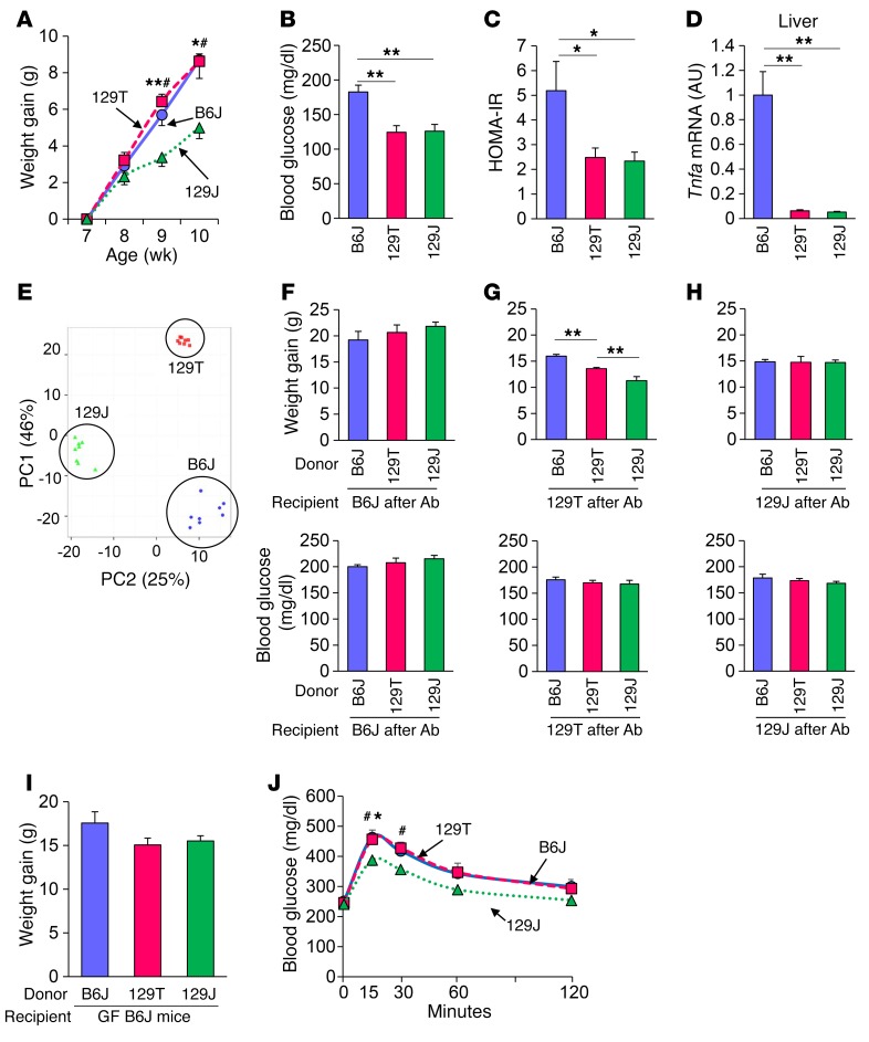
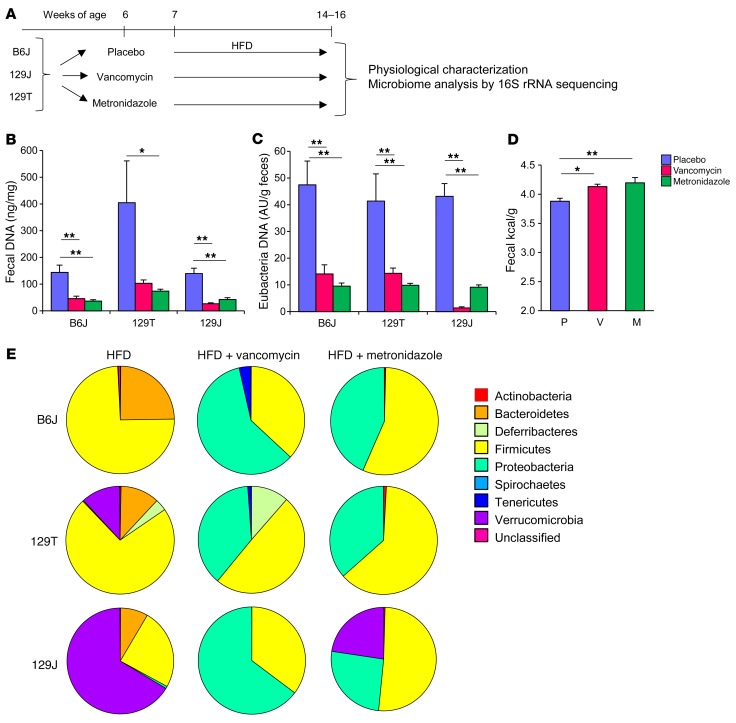
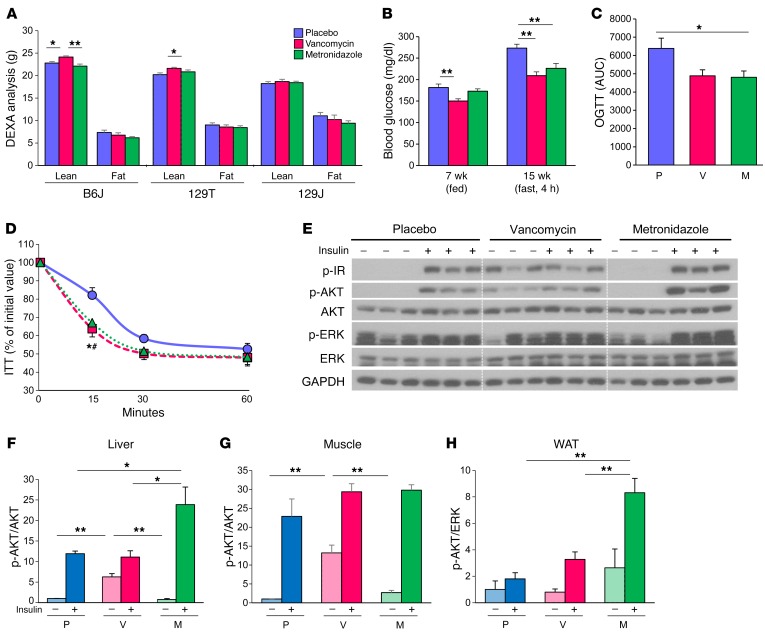
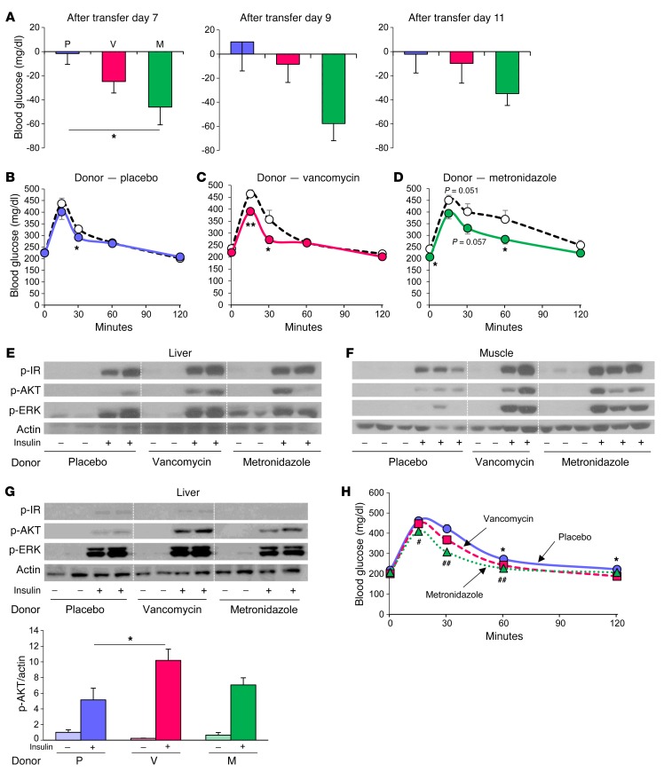
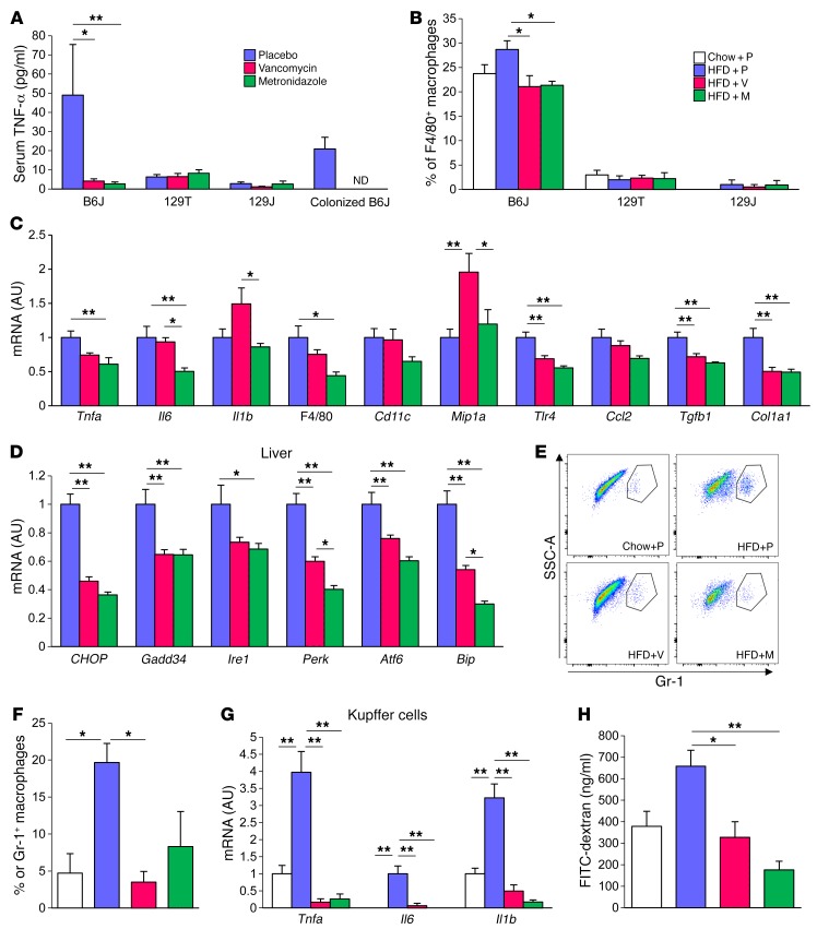
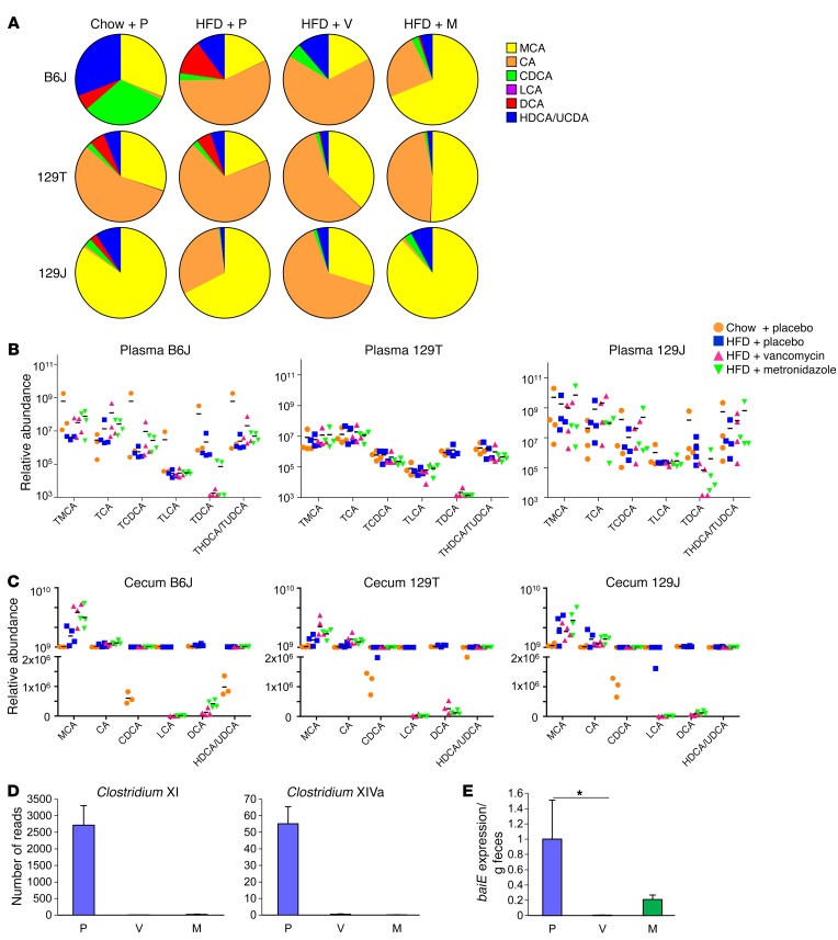
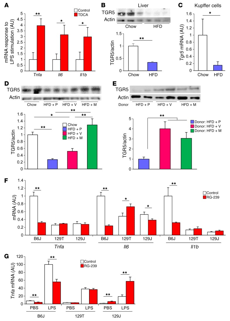
Interactions of diet, gut microbiota, and host genetics play important roles in the development of obesity and insulin resistance. Here, we have investigated the molecular links between gut microbiota, insulin resistance, and glucose metabolism in 3 inbred mouse strains with differing susceptibilities to metabolic syndrome using diet and antibiotic treatment. Antibiotic treatment altered intestinal microbiota, decreased tissue inflammation, improved insulin signaling in basal and stimulated states, and improved glucose metabolism in obesity- and diabetes-prone C57BL/6J mice on a high-fat diet (HFD). Many of these changes were reproduced by the transfer of gut microbiota from antibiotic-treated donors to germ-free or germ-depleted mice. These physiological changes closely correlated with changes in serum bile acids and levels of the antiinflammatory bile acid receptor Takeda G protein-coupled receptor 5 (TGR5) and were partially recapitulated by treatment with a TGR5 agonist. In contrast, antibiotic treatment of HFD-fed, obesity-resistant 129S1 and obesity-prone 129S6 mice did not improve metabolism, despite changes in microbiota and bile acids. These mice also failed to show a reduction in inflammatory gene expression in response to the TGR5 agonist. Thus, changes in bile acid and inflammatory signaling, insulin resistance, and glucose metabolism driven by an HFD can be modified by antibiotic-induced changes in gut microbiota; however, these effects depend on important interactions with the host's genetic background and inflammatory potential.


Figures

Figure 1
Figure 1. Microbiota affect adiposity and glucose metabolism depending on the host genetics.
(A) Weight gain of B6J (circles), 129T (squares), and 129J (triangles) mice on an HFD. #P < 0.05 (129T vs. 129J); *P <0.05 and **P < 0.01 (B6J vs. 129J), by ANOVA, followed by Tukey-Kramer post-hoc. (B) Blood glucose levels in the random fed state at 11 weeks of age (n = 4 per group). (C) Calculated HOMA-IR for B6J, 129T, and 129J mice after 8 weeks on an HFD (n = 8 per group). (D) Tnfa mRNA in liver from mice fed an HFD for 9 weeks as determined by qPCR (n = 8 per group). (E) PCA of fecal 16S rRNA sequencing data for HFD-fed mice of the 3 strains at 16 weeks of age (n = 8 per group). PC1, principle coordinate 1; PC2, principle coordinate 2. (FH) Graphs show weight gain of HFD-fed B6J (F), 129T (G), and 129J (H) mice 10 weeks after undergoing transfer of cecal bacteria from B6J, 129T, and 129J mice (n = 6 per group). Graphs show blood glucose levels (fed) of recipient B6J (F), 129T (G), and 129J (H) mice measured 1 week after bacterial transfer. *P < 0.05 and **P < 0.01, by ANOVA, followed by Tukey-Kramer post-hoc. (I) Weight gain and (J) OGTT for HFD-fed, GF B6J mice colonized with cecal bacteria from B6J (circle), 129T (square), and 129J (triangle) mice measured 2 weeks after transfer (n = 7–10). #P < 0.05 (129T vs. 129J); *P < 0.05 (B6J vs. 129J), by ANOVA, followed by Tukey-Kramer post-hoc.
Figure 2
Figure 2. Antibiotic treatment decreases biomass of the gut bacteria and dramatically modulates microbiota composition.
(A) Schematic overview of the study design for antibiotic modification of the gut microbiota using 3 strains of mice. (B) DNA levels isolated from fecal samples and (C) eubacteria DNA levels measured by qPCR normalized by fecal weight after 1 week of antibiotic treatment (n = 8 per group). (D) Energy content measured by bomb calorimetry of fecal samples from B6J mice after 5 weeks of antibiotic treatment (n = 8). P, placebo; V, vancomycin; M, metronidazole. (E) Representation of bacterial phyla in the fecal microbiota of mice from each group (n = 8) at 14 weeks of age after 8 weeks of antibiotic treatment. *P < 0.05 and **P < 0.01 by ANOVA, followed by Tukey-Kramer post-hoc.
Figure 3
Figure 3. Antibiotic modification of the gut microbiota improves glucose metabolism with improved insulin signaling in HFD-fed B6J mice.
(A) Total fat and lean mass were assessed by DEXA for HFD-fed B6J, 129T, and 129J mice treated with placebo, vancomycin, or metronidazole for 7 weeks (n = 16). (B) Blood glucose levels of HFD-fed B6J mice in the fed (7 weeks old) or 4-hour–fasted state (15 weeks old) (n = 8). (C) AUC of blood glucose levels during an OGTT of 19-week-old HFD-fed B6J mice treated with placebo, vancomycin, or metronidazole (12 weeks on the HFD diet; 13 weeks on antibiotics) (n = 6). (D) ITT of 12-week-old HFD-fed B6J mice treated with placebo, vancomycin, or metronidazole (7 weeks on the HFD; 8 weeks on antibiotics) (n = 16). (E) Western blots for insulin signaling in liver extracts from 16-week-old B6J mice treated with 5 U insulin via the vena cava (9 weeks on the HFD; 10 weeks on antibiotics) n=3. (FH) Quantitation of AKT phosphorylation (p-AKT) normalized by total AKT in liver (F), muscle (G), and adipose tissue (H) extracts from B6J mice treated with placebo, vancomycin, or metronidazole) (n = 3). *P < 0.05 and **P < 0.01, by ANOVA, followed by Tukey-Kramer post-hoc.
Figure 4
Figure 4. Improvement of glucose metabolism by antibiotic-modified bacteria is transferable.
(A) Differences between 4-hour fasting blood glucose levels measured at 1 pm in an HFD-fed B6J mice before and after bacterial transfer (days 7, 9, and 11) from donor HFD-fed B6J mice treated with placebo, vancomycin, or metronidazole for 1 week (n = 6 per group). (BD) OGTT of the HF-fed B6J recipient mice performed before (white circles) and after (solid circles) bacterial transfer (day 11) from mice treated with placebo (B), vancomycin (C), or metronidazole (D) (n = 6). *P < 0.05 and **P < 0.01, by unpaired, 2-tailed t test. (E and F) Western blots for insulin signaling in liver (E) and muscle (F) of the recipient mice. (G) Western blots for insulin signaling in the liver of HFD-fed, GF B6J mice colonized with cecal bacteria from HFD-fed B6J mice treated with placebo, vancomycin, or metronidazole, measured 2 weeks after transfer. Graph shows quantitation of p-AKT protein normalized by actin (n = 4–6). (H) OGTT of HFD-fed, GF B6J mice colonized with cecal bacteria from HFD-fed B6J mice treated with placebo (circles), vancomycin (squares), or metronidazole (triangles) (n = 7–9). *P < 0.05, for placebo versus vancomycin; #P < 0.05 and ##P < 0.01, for placebo versus metronidazole, by ANOVA, followed by Tukey-Kramer post-hoc.
Figure 5
Figure 5. Gut microbiota modification by antibiotics ameliorates diet-induced inflammation in B6J mice.
(A) Serum TNF-α levels of HFD-fed mice treated with placebo, vancomycin, or metronidazole for 9 weeks and of HFD-fed, GF mice colonized with bacteria from placebo-, vancomycin-, or metronidazole-treated (n = 8) mice. (B) Percentage of F4/80+ macrophages in CD11b+CD11c+ cells in lamina propria (n = 5). (C and D) qPCR analysis of inflammatory markers (C) and ER stress markers (D) in the liver (n = 8; 10 weeks of antibiotic treatment). (E) Representative flow cytometric results for Gr-1+ cells in the liver of 8-week-old chow-fed mice and 26-week-old HFD-fed B6J mice (n = 3) (20 weeks on the HFD; 1 week of antibiotic treatment). SSC, side scatter. (F) Percentage of Gr-1+ macrophages in CD11b+F4/80+ Kupffer cells in the liver. (G) qPCR analysis of sorted Kupffer cells (n = 3) and (H) FITC levels in serum of B6J mice after gavage administration of FITC-dextran (n = 3–4). *P < 0.05 and **P < 0.01, by ANOVA, followed by Tukey-Kramer post-hoc.
Figure 6
Figure 6. Antibiotics decrease plasma bile acid metabolites.
(A) Total bile acid composition in the plasma of chow plus placebo–, HFD plus placebo–, HFD plus vancomycin–, and HFD plus metronidazole–treated B6J, 129T, and 129J mice (n = 4) after 4 weeks of treatment. (B and C) Relative abundance of plasma tauro-conjugated bile acids (B) and cecum total bile acids (C) of individual mice from each treatment group. (D) Number of reads for Clostridium XI and Clostridium XIVa clusters (n = 8). (E) qPCR analysis for the baiE gene in feces collected from B6J mice treated for 8 weeks (n = 8). *P < 0.05, by ANOVA, followed by Tukey-Kramer post-hoc.
Figure 7
Figure 7. Modification of the gut microbiota by antibiotics restores bile acid receptor, TGR5 level in the liver.
(A) qPCR for Tnfa, Il6, and Il1b mRNA in peritoneal macrophages collected from mice with or without TDCA treatment (0.4%) for 3 weeks. Cells were stimulated with PBS or LPS (10 ng/ml) for 6 hours. Graph shows the fold change of relative expression levels after the stimulation (n = 5). (B) Western blots for TGR5 in the liver of B6J mice on chow or an HFD for 22 weeks and quantitation of TGR5 protein normalized to actin (n = 4 per group). (C) qPCR for Tgr5 expression in Kupffer cells from mice fed chow or an HFD for 4 to 7 months (n = 8–9). (D) Western blots for TGR5 in the liver of 11-week-old B6J mice on chow or an HFD, with or without antibiotic treatment. Quantitation of TGR5 normalized to actin (n = 4 per group; 4 weeks on the HFD; 5 weeks on antibiotics). (E) Western blots for TGR5 in the liver of HFD-fed, GF B6J mice that received bacterial transfer from placebo-, vancomycin-, or metronidazole-treated mice and quantitation of TGR5 protein normalized to actin (n = 5–12 per group; 19-week-old mice; 10 weeks on the HFD; 2 weeks after colonization). (F) qPCR for Tnfa, Il6, and Il1b expression levels in the liver of mice treated with or without RG-239 (10 mg/kg/d) for 2 weeks (n = 3–5). (G) qPCR for Tnfa expression of peritoneal macrophages collected from mice treated with or without RG-239 and stimulated in vitro with or without 10 ng/ml LPS for 6 hours (n = 4–6). *P < 0.05 and **P < 0.01, by unpaired, 2-tailed t test for (AC and EG) and by ANOVA, followed by Tukey-Kramer post-hoc for (D).

References

    1. Velagapudi VR, et al. The gut microbiota modulates host energy and lipid metabolism in mice. J Lipid Res. 2010;51(5):1101–1112. doi: 10.1194/jlr.M002774. - DOI - PMC - PubMed
    1. Le Chatelier E, et al. Richness of human gut microbiome correlates with metabolic markers. Nature. 2013;500(7464):541–546. doi: 10.1038/nature12506. - DOI - PubMed
    1. Turnbaugh PJ, Ley RE, Mahowald MA, Magrini V, Mardis ER, Gordon JI. An obesity-associated gut microbiome with increased capacity for energy harvest. Nature. 2006;444(7122):1027–1031. doi: 10.1038/nature05414. - DOI - PubMed
    1. Ridaura VK, et al. Gut microbiota from twins discordant for obesity modulate metabolism in mice. Science. 2013;341(6150):1241214. doi: 10.1126/science.1241214. - DOI - PMC - PubMed
    1. Cho I, et al. Antibiotics in early life alter the murine colonic microbiome and adiposity. Nature. 2012;488(7413):621–626. doi: 10.1038/nature11400. - DOI - PMC - PubMed

Publication types

MeSH terms