Review
. 2016 Oct 20;4(4):35.
doi: 10.3390/vaccines4040035.

Tumor-Derived Exosomes and Their Role in Tumor-Induced Immune Suppression

Affiliations
Review

Tumor-Derived Exosomes and Their Role in Tumor-Induced Immune Suppression

Theresa L Whiteside. Vaccines (Basel). .

Abstract

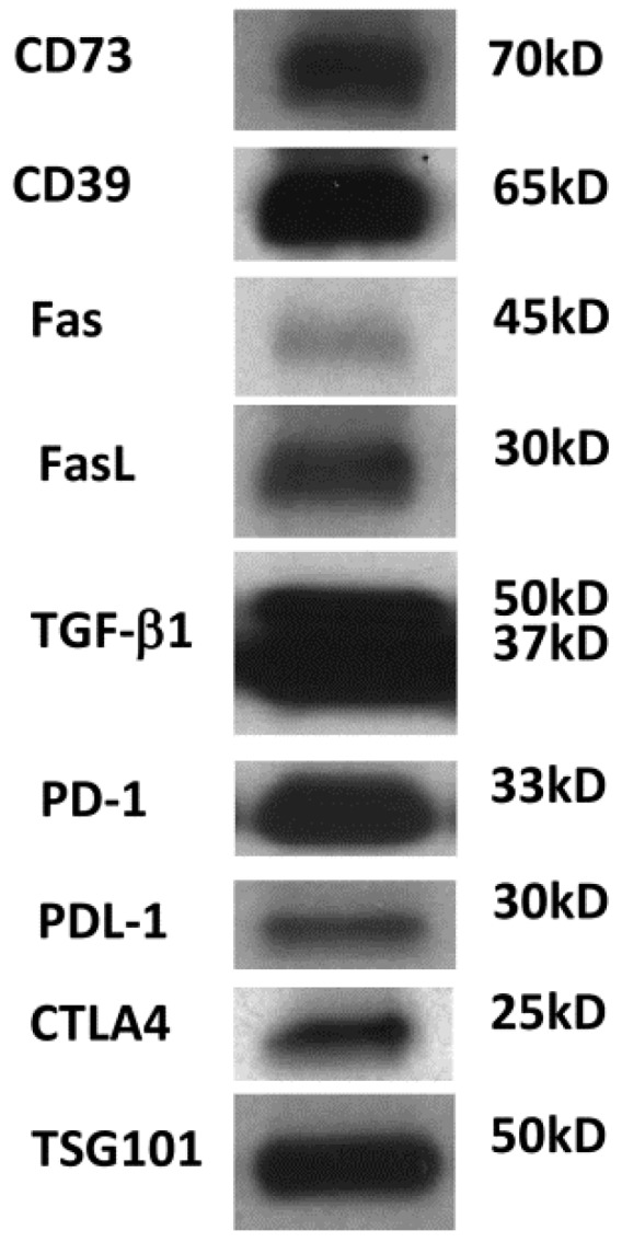
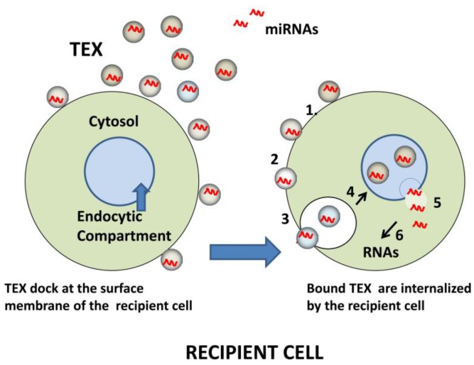
Tumor-derived exosomes (TEX) are emerging as critical components of an intercellular information network between the tumor and the host. The tumor escapes from the host immune system by using a variety of mechanisms designed to impair or eliminate anti-tumor immunity. TEX carrying a cargo of immunoinhibitory molecules and factors represent one such mechanism. TEX, which are present in all body fluids of cancer patients, deliver negative molecular or genetic signals to immune cells re-programming their functions. Although TEX can also stimulate immune activity, in the microenvironments dominated by the tumor, TEX tend to mediate immune suppression thus promoting tumor progression. The TEX content, in part resembling that of the parent cell, may serve as a source of cancer biomarkers. TEX also interfere with immune therapies. A better understanding of TEX and their contribution to cancer progression and cancer patients' response to immune therapies represents a challenging new field of investigation.

Keywords: TEX; biomarkers; cancer; immune suppression; tumor-derived exosomes.


Conflict of interest statement

The author declares no conflicts of interest.

Figures

Figure 1
Figure 1
TEX morphology and composition. In (A), Transmission electron microscopy (TEM) image of TEX isolated by mini-SEC from a supernatant of a head and neck squamous cell carcinoma (HNSCC) cell line, PCI-13. Courtesy of Dr. Sonja Funk. TEX look exactly like other exosomes isolated from cancer patients’ plasma [11]. In (B), a schematic summarizing molecular and genetic contents of TEX surface membrane and lumen is presented. In the lumen, nucleic acids include DNA, mRNA and miRNA; cytosolic protein include various enzymes; soluble factors, such as PGE2 ; cytokines; histones; transport proteins such as ALIX, Rabs, dynamin, LAMPs; cytoskeletal proteins, including actin, tubulin, vimentin and others; oncoproteins; and a variety of signaling molecules, including MAPK, ERK1/2, Rho, catenin, Wnt and many others. The surface membrane of TEX is a lipid-protein bilayer that contains cholesterol, ceramides, sphingomyelins and phospholipids as well as numerous biologically active proteins such as the major histocompatibility complex (MHC) molecules; TAAs; inhibitory ligands such as FasL, TRAIL, PD-L1, TGF-β/LAP; adhesion molecules notably ICAM, EPCAM, CD44, integrins; proteases such as MMPS and CD26; ectonucleotidases engaged in adenosine production, CD39/CD73; transmembrane, receptors such as CXCR4 and c-Met; heat shock proteins (HSPs); and numerous tetraspanins frequently used as “exosome markers.”
Figure 2
Figure 2
Western blot of TEX isolated from the supernatant of human acute myelogenous leukemia (AML) cell line (Kasumi-1) by miniSEC. TEX were concentrated by Viva Spin 300 and loaded onto SDS/PAGE gels always applying 10 µg protein /lane. Western blots were developed as previously described using antibodies specific for the inhibitory proteins [11]. Courtesy of Chang-Sook Hong (University of Pittsburgh Cancer Institute).
Figure 3
Figure 3
Schematic of miRNA transfer to recipient cells by TEX. Initially, TEX, which carry an abundance of adhesion molecules, bind to the surface of a recipient cell (left). Bound TEX (1) may fuse directly with the plasma membrane (2) or be endocytosed (3). The internalized or endocytosed TEX carrying miRNAs can fuse with the membranes delineating the endosomal compartment of the recipient cell (4). TEX are now disrobed and miRNAs are delivered to the cytosol (5), where they interact with complementary mRNAs (6). The result is a change in the transcriptional profile of the recipient cell.

References

    1. Pitt J.M., Marabelle A., Eggermont A., Soria J.C., Kroemer G., Zitvogel L. Targeting the tumor microenvironment: Removing obstruction to anticancer immune responses and immunotherapy. Ann. Oncol. 2016 doi: 10.1093/annonc/mdw168. - DOI - PubMed
    1. Shalapour S., Karin M. Immunity, inflammation, and cancer: An eternal fight between good and evil. J. Clin. Investig. 2015 doi: 10.1172/JCI80007. - DOI - PMC - PubMed
    1. Whiteside T.L. Immune responses to cancer: are they potential biomarkers of prognosis? Front. Oncol. 2013 doi: 10.3389/fonc.2013.00107. - DOI - PMC - PubMed
    1. Whiteside T.L. Immune responses to malignancies. J. Allergy Clin. Immunol. 2010 doi: 10.1016/j.jaci.2009.09.045. - DOI - PMC - PubMed
    1. Abels E.R., Breakefield X.O. Introduction to Extracellular Vesicles: Biogenesis, RNA Cargo Selection, Content, Release, and Uptake. Cell. Mol. Neurobiol. 2016 doi: 10.1007/s10571-016-0366-z. - DOI - PMC - PubMed

LinkOut - more resources