. 2016 Oct 24;11(10):e0161445.
doi: 10.1371/journal.pone.0161445. eCollection 2016.

Tyrosine Binding Protein Sites Regulate the Intracellular Trafficking and Processing of Amyloid Precursor Protein through a Novel Lysosome-Directed Pathway

Affiliations

Tyrosine Binding Protein Sites Regulate the Intracellular Trafficking and Processing of Amyloid Precursor Protein through a Novel Lysosome-Directed Pathway

Joshua H K Tam et al. PLoS One. .

Abstract

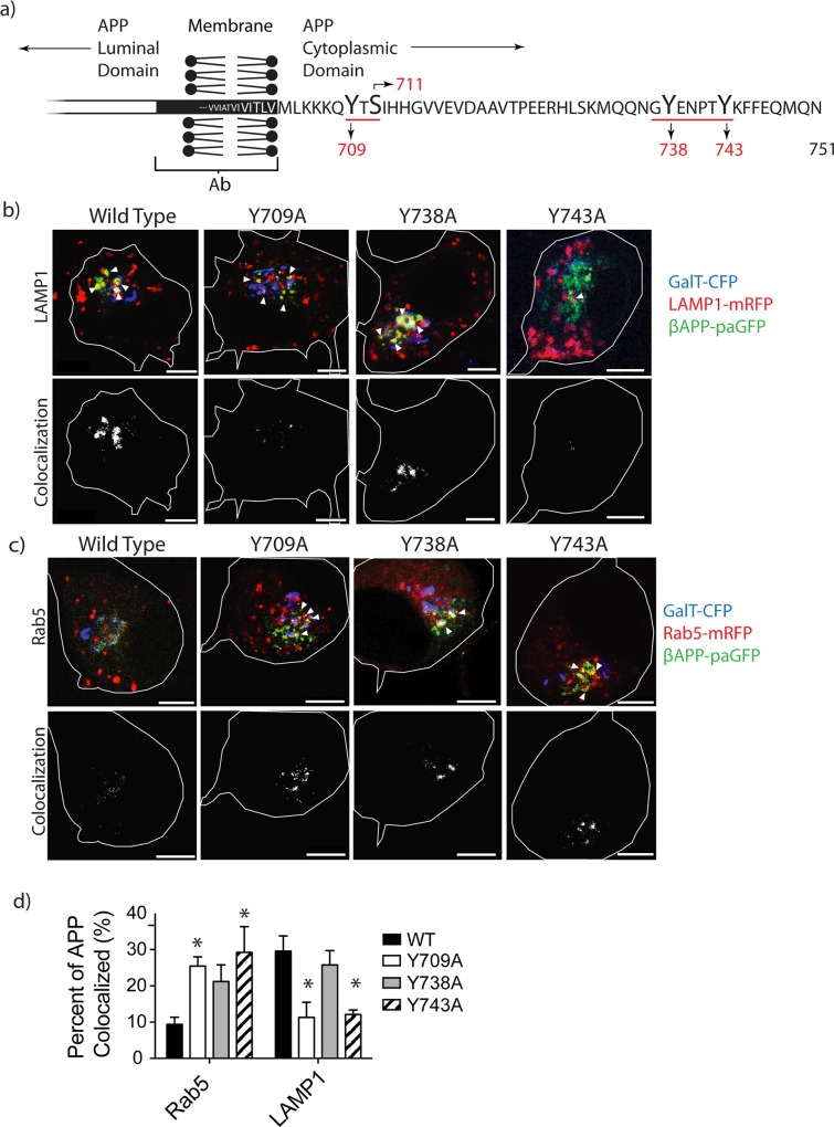
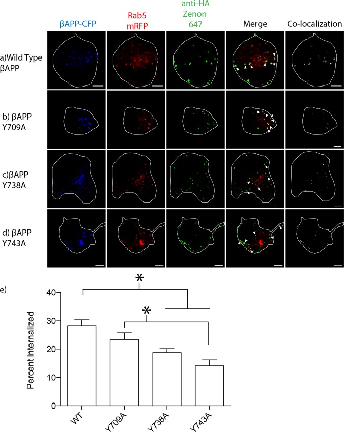
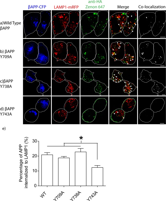
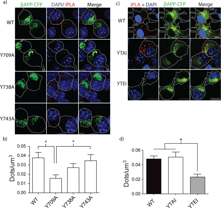
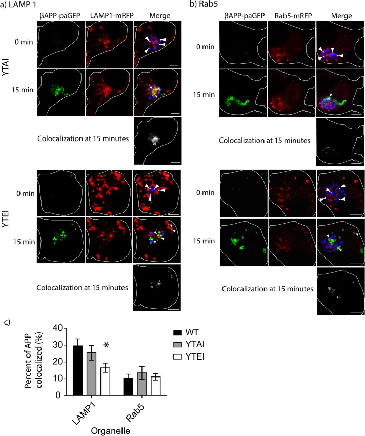
The amyloid hypothesis posits that the production of β-amyloid (Aβ) aggregates leads to neurodegeneration and cognitive decline associated with AD. Aβ is produced by sequential cleavage of the amyloid precursor protein (APP) by β- and γ-secretase. While nascent APP is well known to transit to the endosomal/ lysosomal system via the cell surface, we have recently shown that APP can also traffic to lysosomes intracellularly via its interaction with AP-3. Because AP-3 interacts with cargo protein via interaction with tyrosine motifs, we mutated the three tyrosines motif in the cytoplasmic tail of APP. Here, we show that the YTSI motif interacts with AP-3, and phosphorylation of the serine in this motif disrupts the interaction and decreases APP trafficking to lysosomes. Furthermore, we show that phosphorylation at this motif can decrease the production of neurotoxic Aβ 42. This demonstrates that reducing APP trafficking to lysosomes may be a strategy to reduce Aβ 42 in Alzheimer's disease.


Conflict of interest statement

The authors have declared that no competing interests exist.

Figures

Fig 1
Fig 1. Tyrosine mutations modulate the intracellular trafficking of APP.
a) Depiction of the carboxyl terminal of APP, with APP 751 numbering. The tyrosines and serines studied in this paper are shown, and the tyrosine motifs underlined. SN56 cells were transiently transfected with wild type APP or APP with mutations Y709A, Y738A, or Y743A tagged with paGFP. Each cell was subjected to 15-minutes of sequential imaging. Before each image, the cell was photo-activated within the Golgi (blue). b) Trafficking of APP to lysosomes (LAMP-1) and c) early endosomes (Rab5) was studied. The top panels show representative images from each cell after 15 minutes of photo-activation (Scale bars represent 5μm). The bottom panels depict colocalized pixels. The white border demarcates the edge of the cell and was drawn based on the white light images. Triangles point to colocalized pixels. d) Using a semi-automated method, the APP vesicles and LAMP1 or Rab5 vesicles were selected and the means were plotted using Prism 5.0b. Error bars represent SEM (* = p<0.05).
Fig 2
Fig 2. Tyrosine disrupts internalization into early endosomes.
SN56 cells were transfected with βAPP-CFP (with or without tyrosine mutations) and Rab5-mRFP. The HA-tag on our βAPP-CFP construct was fluorescently labeled using an anti HA-Zenon conjugate. a-d) Representative images of βAPP-CFP, bearing one of the tyrosine mutations, internalized into Rab5-mRFP compartments after 15-minutes. The edge of the cell is shown by the white border, and was drawn based on white-light images. Triangles point to colocalized pixels. Scale bars represent 5μm. e) The percentage of APP co-localized with Rab5 was quantified using Imaris and graphed (* = p<0.05, error bars represent SEM).
Fig 3
Fig 3. Y743A disrupts internalization into lysosomes.
SN56 cells were transfected with βAPP-CFP (with or without tyrosine mutations) and Lamp1-mRFP. The HA-tag was fluorescently labeled using the anti HA-Zenon conjugate. The cells were incubated at 37°C for 15 minutes and fixed and imaged. a-d) Representative images of βAPP-CFP, bearing one of the tyrosine mutations, internalized into LAMP1-mRFP compartments after 15-minutes. The edge of the cell is shown by the white border, and was drawn based on white-light images. Triangles point to colocalized pixels. Scale bars represent 5μm. e) APP co-localized with Lamp1 was quantified using Imaris and graphed (* = p<0.05, error bars represent SEM).
Fig 4
Fig 4. Tyrosine motif mutations affect on APP/AP-3 interaction.
SN56 cells were transfected with plasmids expressing wild type APP or APP with mutations Y709A, Y738A, or Y743A. Cells were fixed and iPLA was performed to detect interaction between APP with AP-3δ. a) Representative images are shown. The white border shows edge of the cell and was drawn based on the white light images. Scale bars represent 5μm. b) The dots per cell was counted using Imaris, normalized to cell volume, and graphed in Prism 5.0b (p<0.05). SN56 cells were transfected with plasmids expressing wild type APP or APP bearing phosphomimetic (S711E) or dephosphomimetic (S711A) mutations. Cells were fixed and iPLA was performed to determine if there was an interaction between APP and AP-3. c) Representative images of APP/AP-3δ interaction. The white border shows edge of the cell and was drawn based on the white light images. Scale bars represent 5μm. d) The number of spots per cell was counted and normalized to cell volume. Error bars represent SEM and * = p<0.05.
Fig 5
Fig 5. S711E disrupts trafficking to lysosomes.
SN56 cells were transfected with plasmids expressing wild type APP, S711E, or S711A. Concomitantly, plasmids expressing LAMP1-mRFP or Rab5-mRFP and GalT-CFP were also transfected. a) Representative images depicting trafficking of APP S711A or S711E to lysosomes (LAMP1-mRFP) after photo-activation in GalT-CFP labeled compartments. b) Representative images showing the delivery of APP S711A or S711E to early endosomes after photo-activation. The edge of the cell is defined by the white line, and was drawn based on the white light images. Scale bars represent 5μm for all images. Triangles with circles denote photo-activation sites at time 0. Triangles alone point to colocalized pixels. c) The percentage of APP colocalized with either LAMP1 or Rab5 was quantified with Imaris. Error bars denote SEM. * = p<0.05.
Fig 6
Fig 6. PMA treatment alters the intracellular trafficking of APP.
SN56 cells transiently transfected with βAPP-paGFP were treated or not treated with 300nM PMA for 1-hour before imaging. Cells were photo-activated in the Golgi (GalT-CFP) for 15 minutes. Video of the live cells was taken during this 15-minute period to follow the trafficking of APP. Frames from the beginning and the end of the time course are shown here for transport to a) lysosomes (LAMP1) and b) early endosomes (Rab5). Far-right panels show colocalized pixels between the βAPP-paGFP and LAMP1-mRFP channels. The edge of the cell is defined by the white line, and was drawn based on the white light images. Triangles alone point to colocalized pixels. Scale bars represent 5μm for all images. c) The amount of APP colocalized with each compartment was quantified using Imaris at the 15-minute time point, and the results were plotted using Prism 5.0b. Error bars represent SEM and * denotes p<0.05.
Fig 7
Fig 7. Staurosporine but not Gö6976 treatment restores trafficking of APP to lysosomes.
SN56 cells were pretreated for 1 hour with staurosporine or Gö6976 for 1 hour before treatment with PMA. Cells were imaged as previously stated. Depicted in a) are representative images of cells treated with PMA, with or without the indicated inhibitors. These images were taken from live cell video of photo-activated cells 15 minutes after the start of imaging. Far-right panels show colocalized pixels between the βAPP-paGFP and LAMP1-mRFP channels. The edge of the cell is defined by the white line, and was drawn based on the white light images. Triangles point to colocalized pixels. Scale bars represent 5μm for all images. b) The amount of APP colocalized with LAMP-mRFP was quantified using Imaris and plotted using Prism. Error bars represent SEM and * denotes p<0.05 as compared to untreated cells and cells treated with staurosporine and PMA.
Fig 8
Fig 8. DCP-LA treatment of SN56 cells diverts APP into early endosome compartments.
Cells were transfected with βAPP-paGFP, GalT-CFP, and a) LAMP1-mRFP or b) Rab5-mRFP. Cells were pre-treated with DCP-LA for one hour before imaging, and photo-activated within the Golgi. Far-right panels show colocalized pixels between the βAPP-paGFP and LAMP1-mRFP channels. The edge of the cell is defined by the white line, and was drawn based on the white light images. Triangles point to colocalized pixels. Scale bars represent 5μm. c) The amount of APP colocalized with each compartment, with or without DCP-LA treatement, was measured using Imaris and was plotted using Prism 5.0b. d) SN56 cells were treated with 1μM staurosproine or 1μM Gö6976 before treatment with DCP-LA. The amount of APP colocalized with LAMP1 was measured using Imaris and plotted using Prism 5.0b. * denotes p<0.05 as compared to untreated. Representative images from the end of the photo-activation period are shown in e). Images in the far-right panel show colocalized pixels between the LAMP1-mrFP and βAPP-paGFP channels. Triangles point to colocalized pixels. Scale bars represent 5μm. f) SN56 cells were transfected with βAPPsw-paGFP and treated with DMSO or DCP-LA. Two other wells of cells were transfected with βAPPsw-paGFP containing either the YTEI and YTAI mutation. The media was collected from the cells and used ELISA to analyze the amount of Aβ42. Error bars in both graphs represent SEM. Results were analyzed by one-way ANOVA with a Tukey’s post hoc test. * denotes p<0.05 as compared to DMSO treated cells.

References

    1. Greenfield JP, Tsai J, Gouras GK, Hai B, Thinakaran G, Checler F, et al. Endoplasmic reticulum and trans-Golgi network generate distinct populations of Alzheimer beta-amyloid peptides. Proc Natl Acad Sci USA. 1999;96: 742–747. - PMC - PubMed
    1. Cirrito JR, Kang J-E, Lee J, Stewart FR, Verges DK, Silverio LM, et al. Endocytosis Is Required for Synaptic Activity-Dependent Release of Amyloid-β In Vivo. Neuron. 2008;58: 42–51. 10.1016/j.neuron.2008.02.003 - DOI - PMC - PubMed
    1. Petanceska SS, Seeger M, Checler F, Gandy S. Mutant presenilin 1 increases the levels of Alzheimer amyloid beta-peptide Abeta42 in late compartments of the constitutive secretory pathway. Journal of Neurochemistry. 2000;74: 1878–1884. - PubMed
    1. Tian Y, Chang JC, Fan EY, Flajolet M, Greengard P. Adaptor complex AP2/PICALM, through interaction with LC3, targets Alzheimer's APP-CTF for terminal degradation via autophagy. Proceedings of the National Academy of Sciences. 2013. 10.1073/pnas.1315110110 - DOI - PMC - PubMed
    1. Thinakaran G, Koo EH. Amyloid Precursor Protein Trafficking, Processing, and Function. Journal of Biological Chemistry. 2008;283: 29615–29619. 10.1074/jbc.R800019200 - DOI - PMC - PubMed

LinkOut - more resources