Scleroderma and dentistry: Two case reports
- PMID: 27776552
- PMCID: PMC5078903
- DOI: 10.1186/s13256-016-1086-1
Scleroderma and dentistry: Two case reports
Abstract
Background: Scleroderma is a chronic connective tissue disorder with unknown etiology. It is characterized by excessive deposition of extracellular matrix in the connective tissues causing vascular disturbances which can result in tissue hypoxia. These changes are manifested as atrophy of the skin and/or mucosa, subcutaneous tissue, muscles, and internal organs. Such changes can be classified into two types, namely, morphea (localized) and diffuse (systemic). Morphea can manifest itself as hemifacial atrophy (Parry-Romberg syndrome) although this remains debatable. Hence, we present a case of morphea, associated with Parry-Romberg syndrome, and a second case with the classical signs of progressive systemic sclerosis.
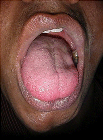
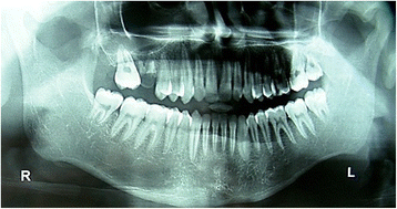
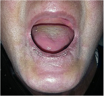
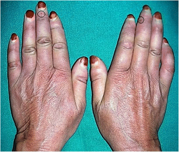
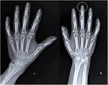
Case presentation: Case one: A 20-year-old man of Dravidian origin presented to our out-patient department with a complaint of facial asymmetry, difficulty in speech, and loss of taste sensation over the last 2 years. There was no history of facial trauma. After physical and radiological investigations, we found gross asymmetry of the left side of his face, a scar on his chin, tongue atrophy, relative microdontia, thinning of the ramus/body of his mandible, and sclerotic lesions on his trunk. Serological investigations were positive for antinuclear antibody for double-stranded deoxyribonucleic acid and mitochondria. A biopsy was suggestive of morphea. Hence, our final diagnosis was mixed morphea with Parry-Romberg syndrome. Case two: A 53-year-old woman of Dravidian origin presented to our out-patient department with a complaint of gradually decreasing mouth opening over the past 7 years. Her medical history was noncontributory. On clinical examination, we found her perioral, neck, and hand skin to be sclerotic. Also, her fingers exhibited bilateral telangiectasia. An oral examination revealed completely edentulous arches as well as xerostomia and candidiasis. Her serological reports were positive for antinuclear antibodies against centromere B, Scl-70, and Ro-52. A hand and wrist radiograph revealed acro-osteolysis of the middle finger on her right hand. Hence, our final diagnosis was progressive systemic sclerosis.
Conclusion: Through this article, we have tried to emphasize the importance of a general examination when diagnosing rare systemic diseases such as scleroderma and the role of the general dentist when caring for such patients, even though they can be quite rare in general practice.
Keywords: CREST syndrome; Localized scleroderma; Morphea; Parry–Romberg syndrome; Raynaud’s phenomenon; Systemic sclerosis.
Figures


Similar articles
-
Progressive hemifacial atrophy or Parry-Romberg syndrome: A pediatric case report.Medwave. 2020 Apr 29;20(3):e7880. doi: 10.5867/medwave.2020.03.7880. Medwave. 2020. PMID: 32428924 English, Spanish.
-
Parry-Romberg syndrome associated with en coup de sabre in a patient from South Sudan - a rare entity from East Africa: a case report.J Med Case Rep. 2019 May 3;13(1):138. doi: 10.1186/s13256-019-2063-2. J Med Case Rep. 2019. PMID: 31046814 Free PMC article.
-
[Neuroretinitis, Parry-Romberg syndrome, and scleroderma].J Fr Ophtalmol. 2005 Oct;28(8):866-70. doi: 10.1016/s0181-5512(05)81008-3. J Fr Ophtalmol. 2005. PMID: 16249769 French.
-
[Parry-Romberg progressive facial hemiatrophy and localized scleroderma. Nosologic and pathogenic problems].J Fr Ophtalmol. 1989;12(3):169-73. J Fr Ophtalmol. 1989. PMID: 2695557 Review. French.
-
Focal Epilepsy in a Teenager With Facial Atrophy and Hair Loss.Semin Pediatr Neurol. 2018 Jul;26:68-73. doi: 10.1016/j.spen.2017.03.009. Epub 2017 Apr 2. Semin Pediatr Neurol. 2018. PMID: 29961525 Review.
Cited by
-
HIV-Associated Systemic Sclerosis: Literature Review and a Rare Case Report.Int J Environ Res Public Health. 2022 Aug 15;19(16):10066. doi: 10.3390/ijerph191610066. Int J Environ Res Public Health. 2022. PMID: 36011703 Free PMC article. Review.
-
Oral manifestations of morphea en plaque: Case report.Ann Med Surg (Lond). 2021 Oct 9;71:102891. doi: 10.1016/j.amsu.2021.102891. eCollection 2021 Nov. Ann Med Surg (Lond). 2021. PMID: 34691443 Free PMC article.
-
Progressive Hemifacial Atrophy and Linear Scleroderma En Coup de Sabre: A Spectrum of the Same Disease?Front Med (Lausanne). 2018 Jan 31;4:258. doi: 10.3389/fmed.2017.00258. eCollection 2017. Front Med (Lausanne). 2018. PMID: 29445726 Free PMC article. Review.
-
Morphea with Oral Mucosa Involvement and Unilateral Nevoid Telangiectasia as an Early Presentation of Morphea: A Case Report and Review of the Literature.J Clin Aesthet Dermatol. 2020 Jan;13(1):38-40. Epub 2020 Jan 1. J Clin Aesthet Dermatol. 2020. PMID: 32082471 Free PMC article.
-
Dacryoadenitis associated with linear scleroderma en coup de sabre: A case report and review of literature.Skin Health Dis. 2024 Apr 22;4(4):e391. doi: 10.1002/ski2.391. eCollection 2024 Aug. Skin Health Dis. 2024. PMID: 39104658 Free PMC article.
References
-
- Goetz RH. The pathology of progressive systemic sclerosis with special reference to changes in the viscera. Clin Proc. 1945;4:337–92.
Publication types
MeSH terms
LinkOut - more resources
Full Text Sources
Other Literature Sources
Medical
Research Materials
Miscellaneous

