

Quantitative computed tomography measurements to evaluate airway disease in chronic obstructive pulmonary disease: Relationship to physiological measurements, clinical index and visual assessment of airway disease
- PMID: 27776670
- PMCID: PMC5310933
- DOI: 10.1016/j.ejrad.2016.09.010
Quantitative computed tomography measurements to evaluate airway disease in chronic obstructive pulmonary disease: Relationship to physiological measurements, clinical index and visual assessment of airway disease
Abstract
Purpose: To correlate currently available quantitative CT measurements for airway disease with physiological indices and the body-mass index, airflow obstruction, dyspnea, and exercise capacity (BODE) index in patients with chronic obstructive pulmonary disease (COPD).
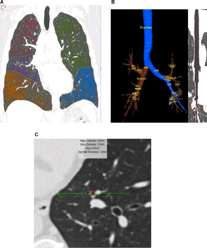
Materials and methods: This study was approved by our institutional review board (IRB number 2778). Written informed consent was obtained from all subjects. The subjects included 188 current and former cigarette smokers from the COPDGene cohort who underwent inspiratory and expiratory CT and also had physiological measurements for the evaluation of airflow limitation, including FEF25-75%, airway resistance (Raw), and specific airway conductance (sGaw). The BODE index was used as the index of clinical symptoms. Quantitative CT measures included % low attenuation areas [% voxels≤950 Hounsfield unit (HU) on inspiratory CT, %LAA-950ins], percent gas trapping (% voxels≤-856HU on expiratory CT, %LAA -856exp), relative inspiratory to expiratory volume change of voxels with attenuation values from -856 to -950HU [Relative Volume Change (RVC)-856 to -950], expiratory to inspiratory ratio of mean lung density (E/I-ratio MLD), Pi10, and airway wall thickness (WT), luminal diameter (LD) and airway wall area percent (WA%) in the segmental, subsegmental and subsubsegmental bronchi on inspiratory CT. Correlation coefficients were calculated between the QCT measurements and physiological measurements in all subjects and in the subjects with mild emphysema (%LAA-950ins <10%). Univariate and multiple variable analysis for the BODE index were also performed. Adjustments were made for age, gender, smoking pack years, FEF25-75%, Raw, and sGaw.
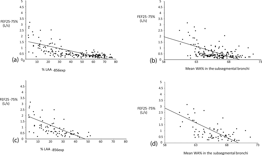
Results: Quantitative CT measurements had significant correlations with physiological indices. Among them, E/I-ratio MLD had the strongest correlations with FEF25-75% (r=-0.648, <0.001) and sGaw (r=-0.624, <0.001) while in the subjects with mild emphysema subsegmental WA% and segmental WA% had the strongest correlation with FEF25-75% (r=-0.669, <0.001) and sGaw (r=-0.638, <0.001), respectively. The multiple variable analyses showed that RVC-856 to -950 was an independent predictor of the BODE index showing the highest R2 (0.468) as an independent variable among the QCT measurements.
Conclusion: Quantitative CT measurements of gas trapping such as E/I-ratio MLD, correlate better with physiological indices for airway disease than those of airway such as WA% or LD. In mild emphysema, however, quantitative CT measurements of airway correlate better with the physiological indices. RVC-856 to -950 is a predictor of the BODE index.
Keywords: Air trapping; Airway disease; CT; Chronic obstructive lung disease (COPD); Quantitative CT.
Copyright © 2016 Elsevier Ireland Ltd. All rights reserved.
Conflict of interest statement
None.
Figures
Similar articles
-
Relationships between airflow obstruction and quantitative CT measurements of emphysema, air trapping, and airways in subjects with and without chronic obstructive pulmonary disease.AJR Am J Roentgenol. 2013 Sep;201(3):W460-70. doi: 10.2214/AJR.12.10102. AJR Am J Roentgenol. 2013. PMID: 23971478 Free PMC article.
-
Relationships between diffusing capacity for carbon monoxide (DLCO), and quantitative computed tomography measurements and visual assessment for chronic obstructive pulmonary disease.Eur J Radiol. 2015 May;84(5):980-5. doi: 10.1016/j.ejrad.2015.01.010. Epub 2015 Jan 22. Eur J Radiol. 2015. PMID: 25704753
-
Relationship between lung function and quantitative computed tomographic parameters of airway remodeling, air trapping, and emphysema in patients with asthma and chronic obstructive pulmonary disease: A single-center study.J Allergy Clin Immunol. 2016 May;137(5):1413-1422.e12. doi: 10.1016/j.jaci.2016.02.001. Epub 2016 Mar 19. J Allergy Clin Immunol. 2016. PMID: 27006248 Free PMC article.
-
Quantitative computed tomography in chronic obstructive pulmonary disease.J Thorac Imaging. 2013 Sep;28(5):284-90. doi: 10.1097/RTI.0b013e318298733c. J Thorac Imaging. 2013. PMID: 23748651 Free PMC article. Review.
-
Imaging Advances in Chronic Obstructive Pulmonary Disease. Insights from the Genetic Epidemiology of Chronic Obstructive Pulmonary Disease (COPDGene) Study.Am J Respir Crit Care Med. 2019 Feb 1;199(3):286-301. doi: 10.1164/rccm.201807-1351SO. Am J Respir Crit Care Med. 2019. PMID: 30304637 Free PMC article. Review.
Cited by
-
Airway remodeling in chronic obstructive pulmonary disease: characteristics and opportunities.Front Med (Lausanne). 2025 Jul 28;12:1556868. doi: 10.3389/fmed.2025.1556868. eCollection 2025. Front Med (Lausanne). 2025. PMID: 40792306 Free PMC article. Review.
-
How anatomical impairments found on CT affect perfusion percentage assessed by SPECT/CT scan?Ann Nucl Med. 2024 Dec;38(12):960-970. doi: 10.1007/s12149-024-01969-7. Epub 2024 Aug 24. Ann Nucl Med. 2024. PMID: 39179897
-
COPD: pulmonary vascular volume associated with cardiac structure and function.Int J Cardiovasc Imaging. 2024 Mar;40(3):579-589. doi: 10.1007/s10554-023-03027-1. Epub 2023 Dec 1. Int J Cardiovasc Imaging. 2024. PMID: 38040946 Free PMC article.
-
Quantitative imaging analysis detects subtle airway abnormalities in symptomatic military deployers.BMC Pulm Med. 2022 Apr 27;22(1):163. doi: 10.1186/s12890-022-01960-w. BMC Pulm Med. 2022. PMID: 35477425 Free PMC article.
-
Quantitative CT lung densitometry as an obstructive marker for the diagnosis of bronchiolitis obliterans in children.PLoS One. 2022 Jul 7;17(7):e0271135. doi: 10.1371/journal.pone.0271135. eCollection 2022. PLoS One. 2022. PMID: 35797398 Free PMC article.
References
-
- Müller NL, Staples CA, Miller RR, Abboud RT. “Density mask”. An objective method to quantitate emphysema using computed tomography. Chest. 1988;94:782–787. - PubMed
-
- Madani A, Zanen J, de Maertelaer V, Gevenois PA. Pulmonary emphysema: radiation dose and section thickness at multidetector CT quantification—comparison with macroscopic and microscopic morphometry. Radiology. 2007;243(1):250–257. - PubMed
-
- Washko GR, Criner GJ, Mohsenifar Z, et al. Computed tomographic-based quantification of emphysema and correlation to pulmonary function and mechanics. COPD. 2008;5(3):177–186. - PubMed
-
- Heussel CP, Herth FJ, Kappes J, et al. Fully automatic quantitative assessment of emphysema in computed tomography: comparison with pulmonary function testing and normal values. Eur. Radiol. 2009;19:2391–2402. - PubMed
-
- D’Anna SE, Asnaghi R, Caramori G, et al. High-resolution computed tomography quantitation of emphysema is correlated with selected lung function values in stable COPD. Respiration. 2012;83(5):383–390. - PubMed
Publication types
MeSH terms
Grants and funding
LinkOut - more resources
Full Text Sources
Other Literature Sources
Medical
Research Materials
