. 2016 Dec 2;291(49):25667-25677.
doi: 10.1074/jbc.M116.759290. Epub 2016 Oct 24.

Structural Analysis of the Catalytic Mechanism and Substrate Specificity of Anabaena Alkaline Invertase InvA Reveals a Novel Glucosidase

Affiliations

Structural Analysis of the Catalytic Mechanism and Substrate Specificity of Anabaena Alkaline Invertase InvA Reveals a Novel Glucosidase

Jin Xie et al. J Biol Chem. .

Abstract

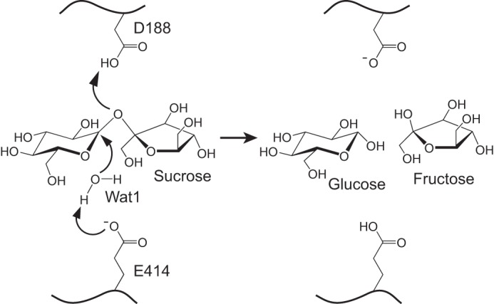
Invertases catalyze the hydrolysis of sucrose to glucose and fructose, thereby playing a key role in primary metabolism and plant development. According to the optimum pH, invertases are classified into acid invertases (Ac-Invs) and alkaline/neutral invertases (A/N-Invs), which share no sequence homology. Compared with Ac-Invs that have been extensively studied, the structure and catalytic mechanism of A/N-Invs remain unknown. Here we report the crystal structures of Anabaena alkaline invertase InvA, which was proposed to be the ancestor of modern plant A/N-Invs. These structures are the first in the GH100 family. InvA exists as a hexamer in both crystal and solution. Each subunit consists of an (α/α)6 barrel core structure in addition to an insertion of three helices. A couple of structures in complex with the substrate or products enabled us to assign the subsites -1 and +1 specifically binding glucose and fructose, respectively. Structural comparison combined with enzymatic assays indicated that Asp-188 and Glu-414 are putative catalytic residues. Further analysis of the substrate binding pocket demonstrated that InvA possesses a stringent substrate specificity toward the α1,2-glycosidic bond of sucrose. Together, we suggest that InvA and homologs represent a novel family of glucosidases.

Keywords: GH100; alkaline/neutral invertases; crystal structure; cyanobacteria; enzyme catalysis; glucosidase; glycoside hydrolase; substrate specificity; sucrose metabolism.


Figures

FIGURE 1.
FIGURE 1.
Overall structure of InvA. A, hexameric structure. Helices are shown as cylinders; sucrose and fructose in the active site are shown as pale cyan and blue sticks, respectively. Two subunits in each dimer are colored in green and yellow, respectively. The six subunits are sequentially labeled as A–F. B, molecular mass determination of InvA by analytical gel filtration chromatography. mAU, milliabsorbance units. C, dimeric structure. A dimer of subunits A and F is shown. D, a close view of InvA monomer. Subunit A in InvA-Suc is used as an example. The core (α/α)6-barrel is shown in green, and the insertion is colored in purple. The sucrose molecule is shown in sticks. E, superposition of InvA against the catalytic domain of tGA, which is shown in gray. F, superposition of truncated InvA (InvA-Suc) against the full-length structure. InvA-Suc and the full-length structure are shown in green and wheat, respectively. A loop at the C terminus of the full-length structure is highlighted in hot pink.
FIGURE 2.
FIGURE 2.
The substrate binding pocket of InvA. Shown are the small molecules binding to the pocket of subunit A (A), subunit C (B), and subunit B (C) of the sucrose-complexed structure InvA-Suc. D, fructose binding to the subsite +1 in the fructose-complexed structure InvA-Fru. E, glucose at the subsite −1 in the glucose-complexed structure InvA-Glc. F, glycerol at the subsite −1 in the full-length structure of InvA. The involved residues are shown in green sticks, the ligands are shown as sticks in different colors, and the water molecules are shown as red spheres. The polar interactions are indicated by dashed lines. The simulated annealing FoFc difference electron density maps of ligands contoured at 3.0 σ are shown as blue mesh.
FIGURE 3.
FIGURE 3.
The optimum pH of InvA. Invertase activity was measured against 100 mm sucrose at pH 5.5–9.5. Three independent experiments were performed, and the S.D. of the mean are shown as error bars.
FIGURE 4.
FIGURE 4.
Catalytic residues of InvA. A, superposition of the active sites of InvA, tGA, and YgjK. The corresponding secondary elements and residues of InvA, tGA, and YgjK are shown in green, gray, and purple, respectively. The catalytic water molecule is displayed as a sphere in a corresponding color. B, the relative activity of the three mutants compared with the wild-type InvA. The error bars represent S.D. from three independent assays.
FIGURE 5.
FIGURE 5.
A putative catalytic mechanism of InvA. Residues Asp-188 and Glu-414 are proposed to be the catalytic acid and base, respectively. Wat1 is the presumed nucleophilic water molecule.
FIGURE 6.
FIGURE 6.
The substrate specificity of InvA. HPLC analysis of the hydrolytic activity of InvA toward 100 mm various sugars. The upper panel is the standard sample of glucose and fructose. The formulas of tested sugars are shown on the right of the corresponding profiles.
FIGURE 7.
FIGURE 7.
Multiple sequence alignment of InvA and homologous A/N-Invs. Secondary structure elements of InvA are shown at the top of alignment. The catalytic residues and substrate binding residues are depicted by red stars and red triangles, respectively. The sequences of InvA and homologs are from the following organisms: Anabaena sp. PCC 7120 (InvA, WP_010995690.1; InvB, CAC85155.1), Fischerella sp. JSC-11 (Fi-A/N-Inv, ZP_08987807.1), Cyanothece sp. PCC 7822 (Cy-A/N-Inv, WP_013325329.1), Arabidopsis thaliana (At-A/N-InvA, NP_176049.1), Daucus carota (Dc-N-Inv, CAA76145.1), Oryza sativa subsp. Japonica (Os-A/N-Inv1, NP_001049936), Triticum aestivum (Ta-A-Inv, CAL26914.1), Ectothiorhodospira sp. PHS-1 (Ec-A/N-Inv, ZP_09695138.1), Halothiobacillus neapolitanus c2 (Hn-A/N-Inv, WP_012823125.1), and Synechocystis sp. PCC 6803 (Sy-A/N-Inv, CAD33848.1).

References

    1. Vargas W. A., and Salerno G. L. (2010) The Cinderella story of sucrose hydrolysis: alkaline/neutral invertases, from cyanobacteria to unforeseen roles in plant cytosol and organelles. Plant Sci. 178, 1–8
    1. Ruan Y.-L., Jin Y., Yang Y.-J., Li G.-J., and Boyer J. S. (2010) Sugar input, metabolism, and signaling mediated by invertase: roles in development, yield potential, and response to drought and heat. Mol. Plant 3, 942–955 - PubMed
    1. Koch K. (2004) Sucrose metabolism: regulatory mechanisms and pivotal roles in sugar sensing and plant development. Curr. Opin. Plant Biol. 7, 235–246 - PubMed
    1. Salerno G. L., and Curatti L. (2003) Origin of sucrose metabolism in higher plants: when, how, and why? Trends Plant Sci. 8, 63–69 - PubMed
    1. Ruan Y.-L. (2014) Sucrose metabolism: gateway to diverse carbon use and sugar signaling. Annu. Rev. Plant Biol. 65, 33–67 - PubMed

LinkOut - more resources