. 2016 Jun 28;7(26):40377-40386.
doi: 10.18632/oncotarget.9750.

Upregulation of SIRT6 predicts poor prognosis and promotes metastasis of non-small cell lung cancer via the ERK1/2/MMP9 pathway

Affiliations

Upregulation of SIRT6 predicts poor prognosis and promotes metastasis of non-small cell lung cancer via the ERK1/2/MMP9 pathway

Lihong Bai et al. Oncotarget. .

Abstract

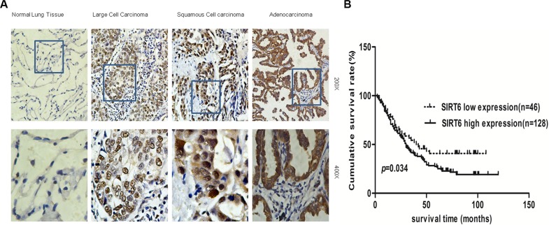
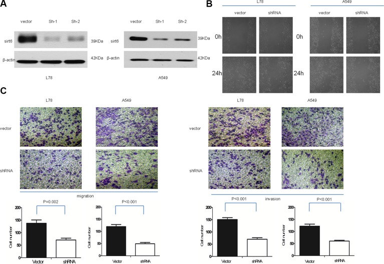
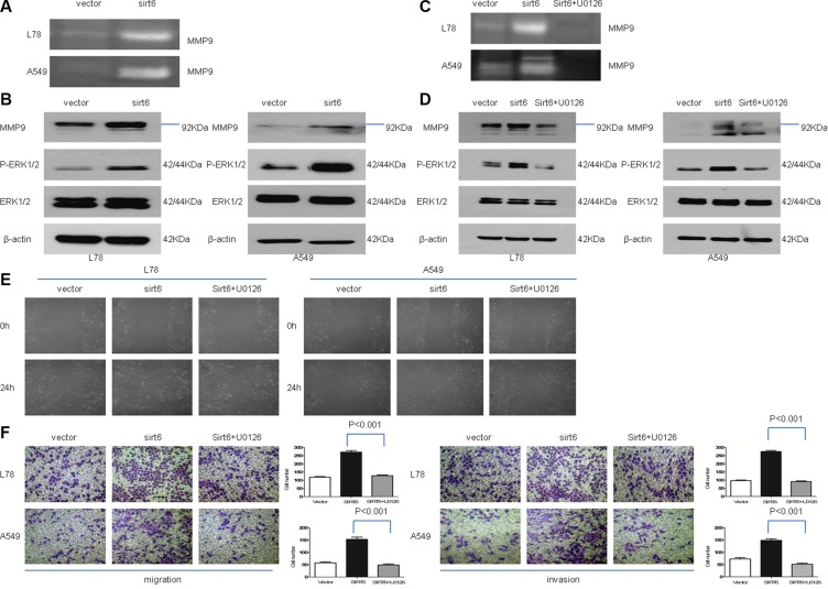
Sirtuin6 (SIRT6), a member of the sirtuins protein family, plays multiple complex roles in cancer. Here, we report that elevated SIRT6 expression was correlated with clinicopathological parameters such as T and N classification in non-small cell lung cancer (NSCLC) patient tumors. SIRT6 overexpression in NSCLC cell lines increased extracellular signal-regulated kinase (p-ERK)1/2 phosphorylation, activated matrix metalloproteinase 9 (MMP9) and promoted tumor cell migration and invasion. Upon treatment with a specific mitogen-activated protein kinase (MEK) 1/2 inhibitor, these effects were abolished. Our results demonstrate SIRT6 upregulation in NSCLC for the first time and suggest a functional role for SIRT6 in promoting migration and invasion through ERK1/2/MMP9 signaling. SIRT6 may serve as a potential therapeutic target in NSCLC and its utility as a prognostic indicator warrants further study.

Keywords: ERK1/2; MMP9; SIRT6; biomarker; non-small cell lung cancer.


Conflict of interest statement

The authors declare no conflicts of interest.

Figures

Figure 1
Figure 1. SIRT6 expression is elevated in both NSCLC cell lines and patient-derived cancer tissues
Western blotting analysis of SIRT6 expression in NSCLC cell lines (A) Western blotting (B) and IHC (C) analysis of SIRT6 expression in primary NSCLC (T) and paired adjacent non-cancerous lung tissue samples (N). Protein levels were normalized with β-actin.
Figure 2
Figure 2. SIRT6 is overexpressed in NSCLC
Representative images of SIRT6 IHC analysis in normal lung tissues and different NSCLC subtypes (A) Survival curves of high and low SIRT6-expressing in NSCLC patients (total n = 174; P = 0.034) (B).
Figure 3
Figure 3. SIRT6 knockdown decreases NSCLC cell migration and invasion
SIRT6 western blotting analysis (A) and wound healing assays (B) in vector-control cells (vector) and SIRT6-shRNA-transduced NSCLC cells (sh1 and sh2); β-actin was used as the loading control for western blotting. Representative micrographs and cell migration and invasion quantification from the transwell migration assay, with and without Matrigel (C) Images represent data from three independent trials with two technical replicates per trial.
Figure 4
Figure 4. Ectopic SIRT6 expression enhances NSCLC cell migration and invasion
SIRT6 western blotting analysis (A) and wound healing assays (B) in A549-vector (vector), A549-SIRT6 (SIRT6), L78-vector (vector) and L78-SIRT6 (SIRT6) cells; β-actin was used as the loading control for western blotting. Representative micrographs and cell migration and invasion quantification from the transwell migration assay, with and without Matrigel (C) Images represent data from three independent trials with two technical replicates per trial.
Figure 5
Figure 5. SIRT6 promotes NSCLC cell migration and invasion through ERK1/2//MMP9
Zymographic analysis of MMP9 activity in conditioned medium from SIRT6-overexpressing NSCLC cells and corresponding vector control cells (A) Western blotting analysis of p-ERK1/2, ERK1/2 and MMP9 in indicated cells (B and D) SIRT6 overexpression increased p-ERK1/2 and MMP9 expression, and treatment with the specific MEK1/2 inhibitor U0126 abolishes these effects. Zymographic analysis of MMP9 activity in conditioned medium from SIRT6-overexpressing cells, with or without U0126 (C) Wound healing assay results showed that stable SIRT6 overexpression promotes cell migration, which is abolished by concomitant treatment with U0126 (E) Migration and invasion assays using a transwell assay system (F) SIRT6-overexpressing cells were treated with U0126 or vehicle alone (without U0126). Representative images and quantification of migration and invasion are shown.

References

    1. Ferlay J, Shin HR, Bray F, Forman D, Mathers C, Parkin DM. Estimates of worldwide burden of cancer in 2008: GLOBOCAN 2008. Int J Cancer. 2010;127:2893–2917. - PubMed
    1. Jemal A, Siegel R, Xu J, Ward E. Cancer statistics, 2010. CA Cancer J Clin. 2010;60:277–300. - PubMed
    1. Jemal A, Siegel R, Ward E, Murray T, Xu J, Thun MJ. CA Cancer J Clin. 2007;57:43–66. - PubMed
    1. Kwon MJ, Seo J, Kim YJ, Kwon MJ, Choi JY, Kim TE, Lee DH, Park S, Shin YK, Han J, Choi YL. Prognostic significance of CD151 overexpression in non-small cell lung cancer. Lung Cancer. 2013;81:109–116. - PubMed
    1. Pei J, Lou Y, Zhong R, Han B. Mmp9 activation triggered by epidermal growth factor induced foxo1 nuclear exclusion in non-small cell lung cancer. Tumour Biol. 2014;35:6673–8. - PubMed

MeSH terms