

Brain structure in pediatric Tourette syndrome
- PMID: 27777415
- PMCID: PMC5405013
- DOI: 10.1038/mp.2016.194
Brain structure in pediatric Tourette syndrome
Erratum in
-
Correction: Brain structure in pediatric Tourette syndrome.Mol Psychiatry. 2020 Nov;25(11):3112. doi: 10.1038/s41380-019-0382-8. Mol Psychiatry. 2020. PMID: 30842575 Free PMC article.
Abstract
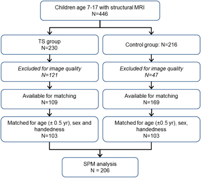
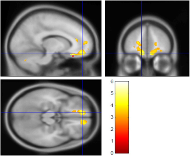
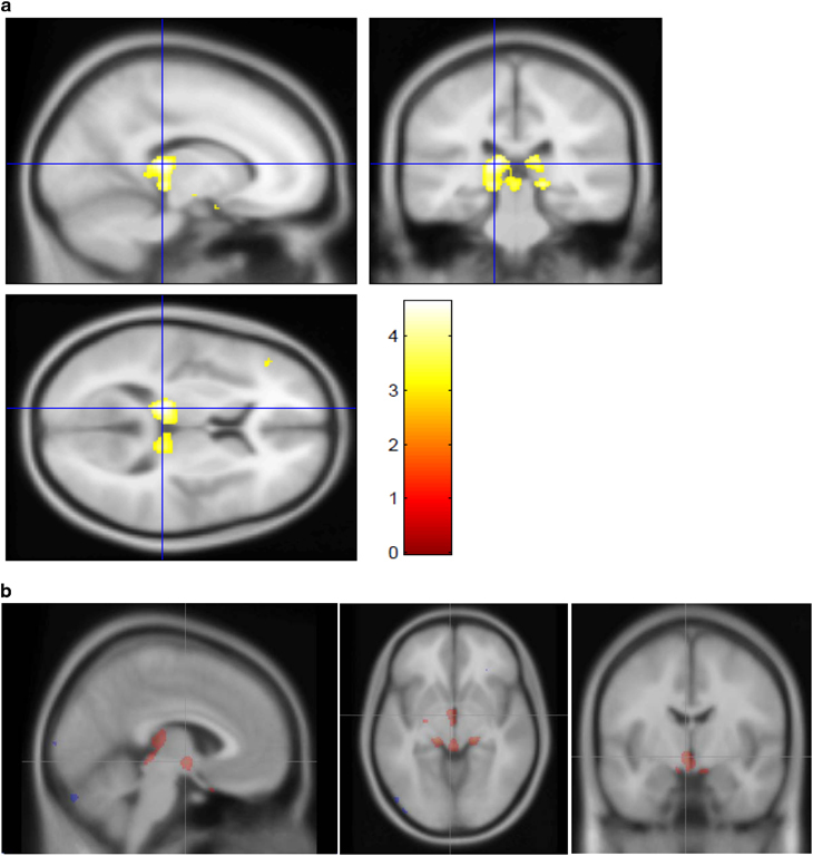
Previous studies of brain structure in Tourette syndrome (TS) have produced mixed results, and most had modest sample sizes. In the present multicenter study, we used structural magnetic resonance imaging (MRI) to compare 103 children and adolescents with TS to a well-matched group of 103 children without tics. We applied voxel-based morphometry methods to test gray matter (GM) and white matter (WM) volume differences between diagnostic groups, accounting for MRI scanner and sequence, age, sex and total GM+WM volume. The TS group demonstrated lower WM volume bilaterally in orbital and medial prefrontal cortex, and greater GM volume in posterior thalamus, hypothalamus and midbrain. These results demonstrate evidence for abnormal brain structure in children and youth with TS, consistent with and extending previous findings, and they point to new target regions and avenues of study in TS. For example, as orbital cortex is reciprocally connected with hypothalamus, structural abnormalities in these regions may relate to abnormal decision making, reinforcement learning or somatic processing in TS.
Conflict of interest statement
The authors declare no conflict of interest.
Figures
Similar articles
-
Structural abnormalities in early Tourette syndrome children: a combined voxel-based morphometry and tract-based spatial statistics study.PLoS One. 2013 Sep 30;8(9):e76105. doi: 10.1371/journal.pone.0076105. eCollection 2013. PLoS One. 2013. PMID: 24098769 Free PMC article.
-
Brain-volume changes in young and middle-aged smokers: a DARTEL-based voxel-based morphometry study.Clin Respir J. 2017 Sep;11(5):621-631. doi: 10.1111/crj.12393. Epub 2015 Oct 13. Clin Respir J. 2017. PMID: 26404024
-
Altered white matter volumes in first-episode depression: Evidence from cross-sectional and longitudinal voxel-based analyses.J Affect Disord. 2019 Feb 15;245:971-977. doi: 10.1016/j.jad.2018.11.085. Epub 2018 Nov 13. J Affect Disord. 2019. PMID: 30699883
-
Structural and functional alterations in the brain gray matter among Tourette syndrome patients: a multimodal meta-analysis of fMRI and VBM studies.J Neurol. 2025 Jan 15;272(2):133. doi: 10.1007/s00415-024-12852-w. J Neurol. 2025. PMID: 39812838 Free PMC article. Review.
-
Cortical and Subcortical Gray Matter Volume in Youths With Conduct Problems: A Meta-analysis.JAMA Psychiatry. 2016 Jan;73(1):64-72. doi: 10.1001/jamapsychiatry.2015.2423. JAMA Psychiatry. 2016. PMID: 26650724 Review.
Cited by
-
Effect of Jian-Pi-Zhi-Dong Decoction on the Amino Acid Neurotransmitters in a Rat Model of Tourette Syndrome and Comorbid Anxiety Disorder.Front Psychiatry. 2020 Jun 5;11:515. doi: 10.3389/fpsyt.2020.00515. eCollection 2020. Front Psychiatry. 2020. PMID: 32581885 Free PMC article.
-
Motor network organization in healthy development and chronic tic disorders.Brain Commun. 2025 Jun 30;7(4):fcaf260. doi: 10.1093/braincomms/fcaf260. eCollection 2025. Brain Commun. 2025. PMID: 40672936 Free PMC article.
-
Altered Functional Connectivity in Resting State Networks in Tourette's Disorder.Front Hum Neurosci. 2018 Sep 18;12:363. doi: 10.3389/fnhum.2018.00363. eCollection 2018. Front Hum Neurosci. 2018. PMID: 30279651 Free PMC article.
-
Behavioral interventions for reducing head motion during MRI scans in children.Neuroimage. 2018 May 1;171:234-245. doi: 10.1016/j.neuroimage.2018.01.023. Epub 2018 Jan 12. Neuroimage. 2018. PMID: 29337280 Free PMC article.
-
Alterations in the microstructure of white matter in children and adolescents with Tourette syndrome measured using tract-based spatial statistics and probabilistic tractography.Cortex. 2018 Jul;104:75-89. doi: 10.1016/j.cortex.2018.04.004. Epub 2018 Apr 12. Cortex. 2018. PMID: 29758375 Free PMC article.
References
-
- American Psychiatric Association Diagnostic and Statistical Manual of Mental Disorders, 5th edn. American Psychiatric Association: Arlington, VA, 2013.
-
- Black KJ, Kompoliti K, Verhagen Metman L. Encyclopedia of Movement Disorders. Elsevier (Academic Press):; 2010. pp. 231–236.
-
- Mink JW. Neurobiology of basal ganglia and Tourette syndrome: basal ganglia circuits and thalamocortical outputs. Adv Neurol. 2006;99:89–98. - PubMed
-
- Martino D, Leckman JF. Tourette Syndrome. Oxford University Press: Oxford; 2013.
Publication types
MeSH terms
Grants and funding
LinkOut - more resources
Full Text Sources
Other Literature Sources
Medical
