. 2016 Jul-Aug;49(4):257-263.
doi: 10.1590/0100-3984.2015.0136.

Abdominal vascular syndromes: characteristic imaging findings

Affiliations

Abdominal vascular syndromes: characteristic imaging findings

Leandro Cardarelli-Leite et al. Radiol Bras. 2016 Jul-Aug.

Abstract

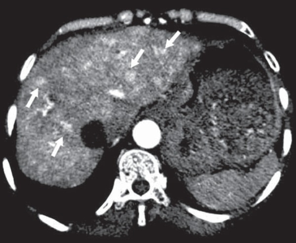
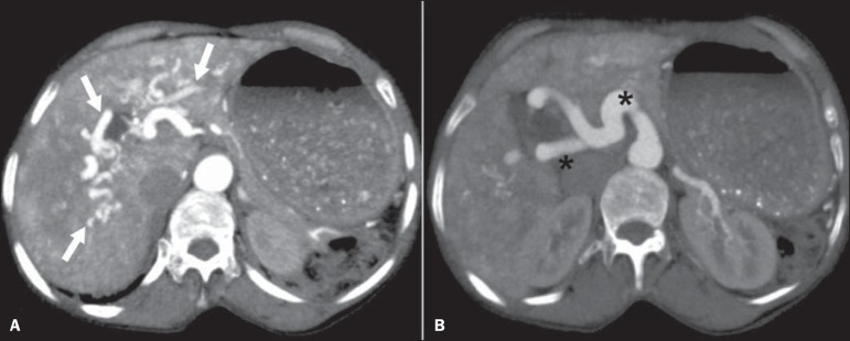
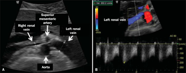
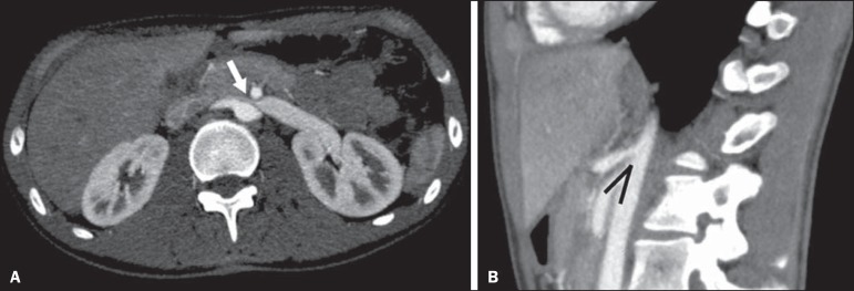
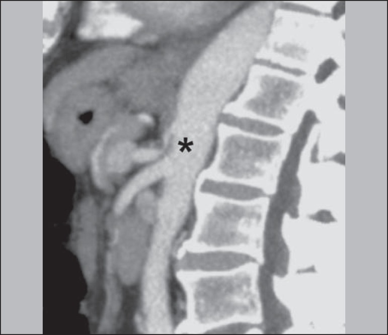
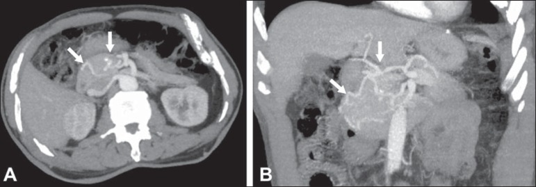
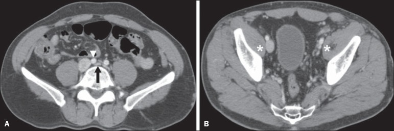
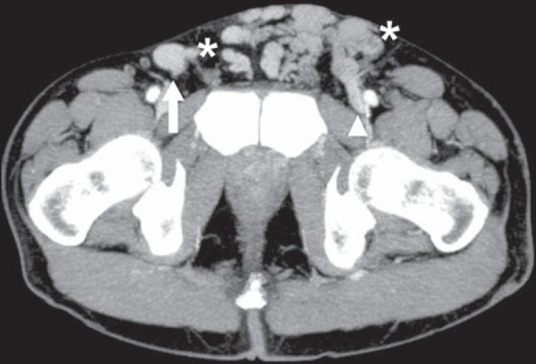
Abdominal vascular syndromes are rare diseases. Although such syndromes vary widely in terms of symptoms and etiologies, certain imaging findings are characteristic. Depending on their etiology, they can be categorized as congenital-including blue rubber bleb nevus syndrome, Klippel-Trenaunay syndrome, and hereditary hemorrhagic telangiectasia (Rendu-Osler-Weber syndrome)-or compressive-including "nutcracker" syndrome, median arcuate ligament syndrome, Cockett syndrome (also known as May-Thurner syndrome), and superior mesenteric artery syndrome. In this article, we aimed to illustrate imaging findings that are characteristic of these syndromes, through studies conducted at our institution, as well as to perform a brief review of the literature on this topic.

As síndromes vasculares abdominais englobam doenças raras, com sintomas e causas diversas, mas com achados de imagem característicos. Dividem-se em dois grupos distintos, a depender de sua origem, que pode ser congênita (síndrome de blue rubber bleb nevus, síndrome de Klippel-Trenaunay e telangiectasia hereditária hemorrágica/Rendu-Osler-Weber) ou compressiva (síndrome do "quebra-nozes", síndrome do ligamento arqueado, síndrome de Cockett ou May-Thurner e síndrome da artéria mesentérica superior). Neste artigo, o nosso objetivo é ilustrar achados de imagens determinantes dessas doenças, por meio de estudos adquiridos em nossa instituição, associando uma breve revisão da literatura.

Keywords: Arteriovenous fistula; Fístula arteriovenosa; Gastrointestinal tract; Hemangioma; Trato gastrintestinal; Trombose venosa; Venous thrombosis.


Figures

Figure 1
Figure 1
Blue rubber bleb nevus syndrome. Axial CT scan showing a calcified hepatic nodule (arrow), suggestive of hemangioma.
Figure 2
Figure 2
Blue rubber bleb nevus syndrome. Sagittal T2-weighted MRI scan showing circumferential thickening of the rectum (asterisk) with high signal intensity.
Figure 3
Figure 3
Klippel-Trenaunay syndrome. Axial maximum intensity projection CT reconstruction showing large caliber gluteal collateral circulation (arrow).
Figure 4
Figure 4
Klippel-Trenaunay syndrome. Axial maximum intensity projection CT reconstruction showing multiple phleboliths distributed throughout the bladder and rectum walls, which are thickened, indicative of cavernous hemangiomas.
Figure 5
Figure 5
Rendu-Osler-Weber syndrome. Axial CT, in the arterial phase, with bone window setting for better visualization of multiple, small hypervascular perfusion disorders (arrows), resulting from intrahepatic shunts.
Figure 6
Figure 6
Rendu-Osler-Weber syndrome. Axial maximum intensity projection CT reconstructions. A: Ectasia and intrahepatic arterial sinuosity characteristic of arterial malformations (arrows). B: Accentuated increase in caliber throughout the hepatic artery and main branches (asterisk).
Figure 7
Figure 7
Nutcracker syndrome. A: B-mode ultrasound showing left renal artery stenosis between the superior mesenteric artery and the aorta, with pre-stenotic ectasia. B: Doppler ultrasound showing an accentuated increase in velocity in the left renal vein immediately downstream of the stenosis.
Figure 8
Figure 8
Nutcracker syndrome. A: Axial CT identifying stenosis in the left renal artery, between the superior mesenteric artery and the aorta (arrow), with pre-stenotic ectasia. B: Sagittal CT showing a reduction in the aortomesenteric angle (arrow point).
Figure 9
Figure 9
Median arcuate ligament syndrome. Sagittal CT showing a "hooked" aspect (trajectory flattening) of the celiac trunk (asterisk), with mild post-stenotic ectasia.
Figure 10
Figure 10
Median arcuate ligament syndrome. Axial (A) and coronal (B) maximum intensity projection CT reconstructions showing prominence (arrows) of the pancreaticoduodenal arcade (collateral circulation between the celiac trunk and the superior mesenteric artery).
Figure 11
Figure 11
Cockett syndrome or May-Thurner syndrome. Axial CT. A: Compression of the left common iliac vein (arrow) by the right common iliac artery (arrowhead). B: Asymmetry of the external iliac veins (asterisk), with diffuse tapering on the left (chronic thrombosis) and ectasia on the right (resulting from increased pelvic collateral flow).
Figure 12
Figure 12
Cockett syndrome or May-Thurner syndrome. Axial CT showing suprapubic venous collateral circulation (asterisk), which creates communication between the right and left common femoral veins (arrow and arrowhead, respectively).
Figure 13
Figure 13
Superior mesenteric artery syndrome. Axial CT showing compression of the third part of the duodenum (arrow) between the superior mesenteric artery and the aorta, with consequent upstream dilatation (asterisk).
Figure 14
Figure 14
Superior mesenteric artery syndrome. A: Sagittal CT showing reduction of the aortomesenteric angle (arrow point) with reduction of the duodenal caliber at that level (arrow). B: Coronal CT showing gastroduodenal dilatation upstream of the compression.

Similar articles

Cited by

References

    1. Nascimento JHR, Soder RB, Epifanio M, et al. Accuracy of computer-aided ultrasound as compared with magnetic resonance imaging in the evaluation of nonalcoholic fatty liver disease in obese and eutrophic adolescents. Radiol Bras. 2015;48:225–232. - PMC - PubMed
    1. Vermelho MBF, Correia AS, Michailowsky TCA, et al. Abdominal alterations in disseminated paracoccidioidomycosis: computed tomography findings. Radiol Bras. 2015;48:81–85. - PMC - PubMed
    1. Szejnfeld D, Nunes TF, Fornazari VAV, et al. Transcatheter arterial embolization for unresectable symptomatic giant hepatic hemangiomas: single-center experience using a lipiodol-ethanol mixture. Radiol Bras. 2015;48:154–157. - PMC - PubMed
    1. Bormann RL, Rocha EL, Kierszenbaum ML, et al. The role of gadoxetic acid as a paramagnetic contrast medium in the characterization and detection of focal liver lesions: a review. Radiol Bras. 2015;48:43–51. - PMC - PubMed
    1. Barros RHO, Penachim TJ, Martins DL, et al. Multidetector computed tomography in the preoperative staging of gastric adenocarcinoma. Radiol Bras. 2015;48:74–80. - PMC - PubMed

LinkOut - more resources