Ewing Sarcoma in the Right Ventricle
- PMID: 27777536
- PMCID: PMC5067046
- DOI: 10.14503/THIJ-15-5330
Ewing Sarcoma in the Right Ventricle
Abstract
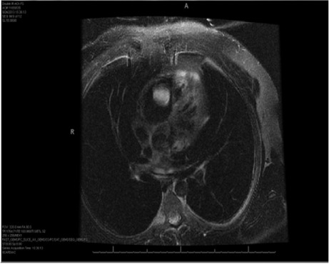
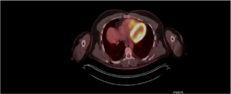
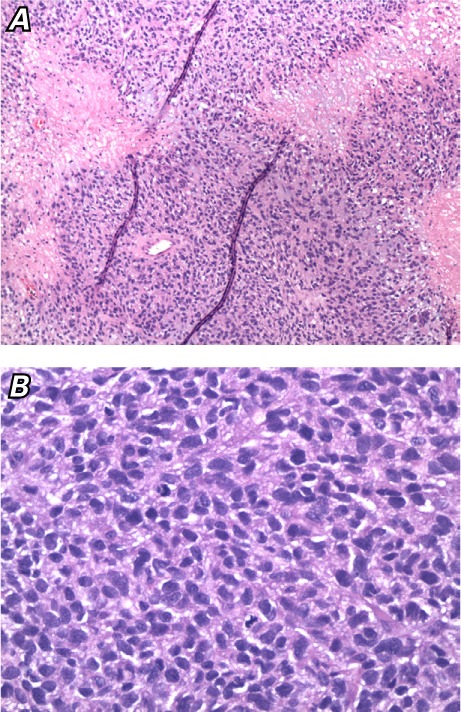
Ewing sarcoma is the second most prevalent malignant primary bone tumor but constitutes only a small proportion of cardiac metastases. We present a case of asymptomatic Ewing sarcoma metastatic to the right ventricle. A 36-year-old man presented for evaluation and resection of a pedunculated right ventricular cardiac tumor. Three years before, he had been diagnosed with translocation-negative Ewing sarcoma, for which he had undergone chemotherapy and amputation of the left leg below the knee. We resected the right ventricular tumor. Analysis of the resected mass supported the diagnosis of metastatic Ewing sarcoma. Postoperative transthoracic echocardiograms showed normal biventricular size and function. One year later, the patient had no recurrence of the sarcoma. In addition to discussing this case, we review the relevant medical literature.
Keywords: Heart neoplasms/secondary/surgery; heart ventricles/surgery; neoplasm invasiveness; oncogene proteins, fusion; sarcoma, Ewing/genetics/pathology/surgery; treatment outcome.
Figures
Similar articles
-
A Case of Ewing Sarcoma Presenting With Cardiac Metastasis.World J Pediatr Congenit Heart Surg. 2019 Sep;10(5):632-634. doi: 10.1177/2150135119846047. World J Pediatr Congenit Heart Surg. 2019. PMID: 31496409
-
Extraskeletal Ewing's sarcoma of primary cardiac origin.Pediatr Cardiol. 1994 Jul-Aug;15(4):207-8. doi: 10.1007/BF00800678. Pediatr Cardiol. 1994. PMID: 7991441
-
"Second" Primary Cardiac Sarcoma in a Patient With Ewing Sarcoma. Always Expect The Unexpected.Ann Thorac Surg. 2017 Feb;103(2):e131-e133. doi: 10.1016/j.athoracsur.2016.07.063. Ann Thorac Surg. 2017. PMID: 28109371
-
Histologic Features and Prognosis of Spinal Intradural Extramedullary Ewing Sarcoma: Case Report, Literature Review, and Analysis of Prognosis.World Neurosurg. 2018 Jul;115:448-452.e2. doi: 10.1016/j.wneu.2018.04.015. Epub 2018 Apr 11. World Neurosurg. 2018. PMID: 29654955 Review.
-
Primary Ewing sarcoma of the larynx with distant metastasis: a case report and review of the literature.Curr Oncol. 2019 Aug;26(4):e574-e577. doi: 10.3747/co.26.5001. Epub 2019 Aug 1. Curr Oncol. 2019. PMID: 31548827 Free PMC article. Review.
Cited by
-
Atrial Thrombus Mimicking Ewing's Sarcoma.CASE (Phila). 2019 Sep 12;3(6):280-283. doi: 10.1016/j.case.2019.07.006. eCollection 2019 Dec. CASE (Phila). 2019. PMID: 32002485 Free PMC article.
-
Right Ventricular Metastasis of Ewing Sarcoma Treated Through Surgical Resection.JACC Case Rep. 2024 Dec 4;29(23):102654. doi: 10.1016/j.jaccas.2024.102654. eCollection 2024 Dec 4. JACC Case Rep. 2024. PMID: 39691334 Free PMC article.
-
Adult-onset primary Ewing's sarcoma of the right atrium: a case report.Surg Case Rep. 2019 Nov 6;5(1):171. doi: 10.1186/s40792-019-0727-1. Surg Case Rep. 2019. PMID: 31696353 Free PMC article.
References
-
- Pinder M, Charafeddine A, Parnell AS, DiBardino DJ, Knudson JD. Osteosarcoma with cardiac metastasis in a 22-year-old man: a case report and review of cardiac tumors. Congenit Heart Dis. 2014;9(5):E147–52. - PubMed
-
- Klatt EC, Heitz DR. Cardiac metastases. Cancer. 1990;65(6):1456–9. - PubMed
-
- Bernstein M, Kovar H, Paulussen M, Randall RL, Schuck A, Teot LA, Juergens H. Ewing's sarcoma family of tumors: current management. Oncologist. 2006;11(5):503–19. - PubMed
-
- Coccia P, Ruggiero A, Rufini V, Maurizi P, Attina G, Marano R et al. Cardiac metastases of Ewing sarcoma detected by 18F-FDG PET/CT. J Pediatr Hematol Oncol. 2012;34(3):236–8. - PubMed
Publication types
MeSH terms
LinkOut - more resources
Full Text Sources
Other Literature Sources
Medical