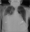
Giant Left Atrium Causing Dysphagia
- PMID: 27777540
- PMCID: PMC5067050
- DOI: 10.14503/THIJ-15-5682
Giant Left Atrium Causing Dysphagia
Figures


References
-
- Lang RM, Badano LP, Mor-Avi V, Afilalo J, Armstrong A, Ernande L et al. Recommendations for cardiac chamber quantification by echocardiography in adults: an update from the American Society of Echocardiography and the European Association of Cardiovascular Imaging. J Am Soc Echocardiogr. 2015;28(1):1–39.e14. - PubMed
-
- Hurst JW. Memories of patients with a giant left atrium. Circulation. 2001;104(22):2630–1. - PubMed
-
- Kawazoe K, Beppu S, Takahara Y, Nakajima N, Tanaka K, Ichihashi K et al. Surgical treatment of giant left atrium combined with mitral valvular disease. Plication procedure for reduction of compression to the left ventricle, bronchus, and pulmonary parenchyma. J Thorac Cardiovasc Surg. 1983;85(6):885–92. - PubMed
-
- Isomura T, Hisatomi K, Hirano A, Maruyama H, Kosuga K, Ohishi K et al. Left atrial plication and mitral valve replacement for giant left atrium accompanying mitral lesion. J Card Surg. 1993;8(3):365–70. - PubMed
-
- Apostolakis E, Shuhaiber JH. The surgical management of giant left atrium. Eur J Cardiothorac Surg. 2008;33(2):182–90. - PubMed
Publication types
MeSH terms
LinkOut - more resources
Full Text Sources
Other Literature Sources
Medical

