

Burn injuries and soft tissue traumas complicated by mucormycosis infection: a report of six cases and review of the literature
- PMID: 27777549
- PMCID: PMC5068897
Burn injuries and soft tissue traumas complicated by mucormycosis infection: a report of six cases and review of the literature
Abstract
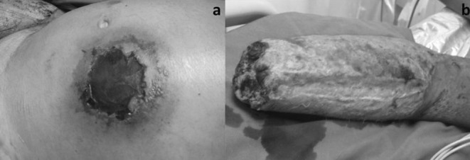
Mucor fungus infection is a rare opportunistic infection, rapidly progressive and often fatal in immunocompromised patients, or in patients with chronic debilitating diseases. We report six cases of trauma patients with mucormycosis. Three had severe thermal burns, one of them with a medical history of diabetes mellitus. The other three patients suffered from severe soft tissue injuries caused by traffic accidents. In all cases there had been spontaneous exposure and contact of the wounds with soil. During hospitalization, fungi cultures and/or biopsies of all wounds were performed and all resulted positive. The patients were treated with Amphotericin B (AmB) and surgical debridement. Two of them died and the other four were fully healed and discharged. Mucormycosis should be considered in any case of aggressive skin tissue necrosis with a history of soiled wounds. We suggest that mucormycosis is treated by intravenous and local administration of AmB, extensive and repeated debridement and cautious coverage of the wound. The plastic surgeon must wait for negative swab cultures and biopsies before covering the defects with skin grafts or flaps. Reconstruction may be challenging, depending on the extent, depth, location and special indications of the affected site and the donor site availability.
L’infection fongique (mucor mycose) est une infection rare, opportuniste, d’aggravation rapide et d’évolution souvent fatale chez les patients immunodéprimés ou porteurs d’affections chroniques fragilisantes. Nous rapportons 6 cas de traumatismes avec mucor mycoses; 3 d’entre eux présentaient des brûlures thermiques sévères: l’un était atteint de diabète, les 3 autres patients souffraient de graves lésions des parties molles secondaires à des accidents de circulation; dans tous les cas, il s’agissait de plaies largement ouvertes et souillées; pendant l’hospitalisation les cultures ou les biopsies objectivaient la présence fongique; les patients furent traités par Amphotiricine B (AmB) et détersion chirurgicale; 2 patients moururent et les 4 autres évoluèrent vers la cicatrisation complète. La mucor mycose doit être évoquée devant tout cas de lésions cutanées profondes et nécrotiques avec notion de souillure. Nous suggérons que la mucor mycose soit traitée par administration intraveineuse et locale d’AmB avec détersion répétée, large et protection prudente de la plaie. Le chirurgien plastique doit attendre la négativité des cultures après écouvillonnage et biopsie avant de couvrir les pertes de substance par greffe ou lambeau. La reconstruction est un challenge qui dépend de l’étendue, de la profondeur, de la localisation et des particularités de la lésion ainsi que de la disponibilité de zone donneuse.
Keywords: brûlures; infection fongique; mucor mycose; traumatismes.
Figures


References
-
- Lehrer RL, Howard DH, Sypherd PS et al. Mucormycosis. Ann Int Med. 1980;93:93–108.
-
- Tang D, Wang W. Successful cure of an extensive burn injury complicated with mucor wound sepsis. Burns. 1998;24:72–73. - PubMed
-
- Becker WK, Cioffi WG Jr, McManus AT et al. Fungal burn wound infection. A ten-year experience. Arch Surg. 1991;126:44–48. - PubMed
LinkOut - more resources
Full Text Sources