Review
. 2016 Oct 10:7:387.
doi: 10.3389/fimmu.2016.00387. eCollection 2016.

Artery Tertiary Lymphoid Organs: Powerhouses of Atherosclerosis Immunity

Affiliations
Review

Artery Tertiary Lymphoid Organs: Powerhouses of Atherosclerosis Immunity

Changjun Yin et al. Front Immunol. .

Abstract

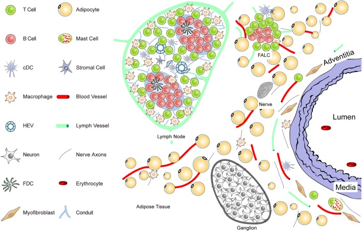
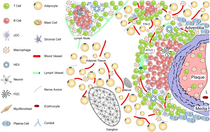
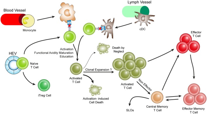
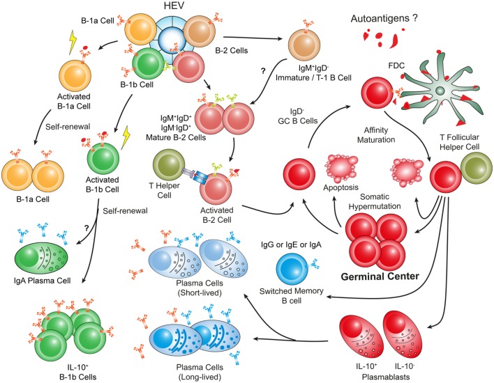
Artery tertiary lymphoid organs (ATLOs) are atherosclerosis-associated lymphoid aggregates with varying degrees of complexity ranging from small T/B-cell clusters to well-structured lymph node-like though unencapsulated lymphoid tissues. ATLOs arise in the connective tissue that surrounds diseased arteries, i.e., the adventitia. ATLOs have been identified in aged atherosclerosis-prone hyperlipidemic apolipoprotein E-deficient (ApoE-/-) mice: they are organized into distinct immune cell compartments, including separate T-cell areas, activated B-cell follicles, and plasma cell niches. Analyses of ATLO immune cell subsets indicate antigen-specific T- and B-cell immune reactions within the atherosclerotic arterial wall adventitia. Moreover, ATLOs harbor innate immune cells, including a large component of inflammatory macrophages, B-1 cells, and an aberrant set of antigen-presenting cells. There is marked neoangiogenesis, irregular lymphangiogenesis, neoformation of high endothelial venules, and de novo synthesis of lymph node-like conduits. Molecular mechanisms of ATLO formation remain to be identified though media vascular smooth muscle cells may adopt features of lymphoid tissue organizer-like cells by expressing lymphorganogenic chemokines, i.e., CXCL13 and CCL21. Although these data are consistent with the view that ATLOs participate in primary T- and B-cell responses against elusive atherosclerosis-specific autoantigens, their specific protective or disease-promoting roles remain to be identified. In this review, we discuss what is currently known about ATLOs and their potential impact on atherosclerosis and make attempts to define challenges ahead.

Keywords: adventitia; aging; artery tertiary lymphoid organs; atherosclerosis; autoimmune responses.


Figures

Figure 1
Figure 1
Anatomy of the normal aorta adventitia and adjacent tissues. The normal adventitial layer of the mouse aorta constitutively contains T-cells (but no B-cells), tissue macrophages, cDCs, stromal cells or myofibroblasts, and mast cells. The adjacent tissues including the periaortic adipose tissue harbors fat-associated lymphoid clusters (FALCs) in wild-type mice. FALCs represent T/B-cell aggregates of various sizes that are exclusively observed in adipose tissue. In the adventitia, axons of both the sensory and the sympathetic nervous systems have been identified as well as vasa vasorum and lymph vessels. Ganglia of the peripheral nervous system are embedded in the adipose tissue, and their axons reach the external lamina that separates the adventitia from the media layer.
Figure 2
Figure 2
Anatomy of the diseased aorta adventitia in aged hyperlipidemic mice. Aged hyperlipidemic mice develop ATLOs in the adventitia layer of the aorta selectively adjacent to atherosclerotic plaques with a preference for the abdominal aorta segment. ATLOs range from small T/B-cell aggregates to well-structured TLOs with separate T- and B-cell areas. The most advanced forms of ATLOs contain B-cell follicles including FDCs, newly formed conduits, HEVs, and aberrant lymph vessels (see Figure 4).
Figure 3
Figure 3
Hypothetical choreography for antigen-dependent ATLO T-cell response pathways. ATLOs are powerhouses of T-cell immunity. They promote T-cell recruitment and suppress T-cell egress allowing for extended T/DC interactions, activation, proliferation, and education of T-cells. ATLOs also mediate conversion of naïve CD4+ T-cells into iTreg cells. This schematic representation has been modified from Hu et al. reported earlier [see Ref. (67) for detailed data].
Figure 4
Figure 4
Hypothetical choreography of antigen-dependent ATLO B-cell response pathways. Advanced ATLOs are powerhouses of B-cell immunity. ATLO B-cell responses are two-pronged in nature containing a comprehensive B-1 and a multi-faceted B-2 maturation pathway. Although similarities of B-cell immunity in ATLOs and SLOs are apparent, differences include a large PC component and a distinct B-1 B cell compartment which differs from that in the peritoneal cavity. This schematic figure has been modified from Srikakulapu et al. (68) [see Ref. (68) for detailed data].

References

    1. Ait-Oufella H, Sage AP, Mallat Z, Tedgui A. Adaptive (T and B cells) immunity and control by dendritic cells in atherosclerosis. Circ Res (2014) 114:1640–60.10.1161/CIRCRESAHA.114.302761 - DOI - PubMed
    1. Glass CK, Witztum JL. Atherosclerosis. the road ahead. Cell (2001) 104:503–16.10.1016/S0092-8674(01)00238-0 - DOI - PubMed
    1. Hansson GK, Hermansson A. The immune system in atherosclerosis. Nat Immunol (2011) 12:204–12.10.1038/ni.2001 - DOI - PubMed
    1. Libby P. Mechanisms of acute coronary syndromes and their implications for therapy. N Engl J Med (2013) 368:2004–13.10.1056/NEJMra1216063 - DOI - PubMed
    1. Swirski FK, Nahrendorf M. Leukocyte behavior in atherosclerosis, myocardial infarction, and heart failure. Science (2013) 339:161–6.10.1126/science.1230719 - DOI - PMC - PubMed