Review
. 2016:2016:6032306.
doi: 10.1155/2016/6032306. Epub 2016 Sep 29.

Targeting Glial Mitochondrial Function for Protection from Cerebral Ischemia: Relevance, Mechanisms, and the Role of MicroRNAs

Affiliations
Review

Targeting Glial Mitochondrial Function for Protection from Cerebral Ischemia: Relevance, Mechanisms, and the Role of MicroRNAs

Le Li et al. Oxid Med Cell Longev. 2016.

Abstract

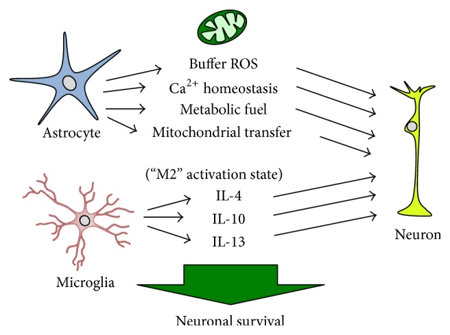
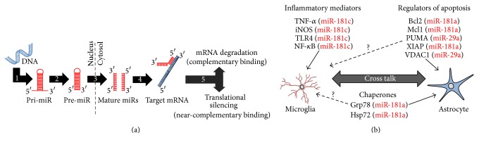
Astrocytes and microglia play crucial roles in the response to cerebral ischemia and are effective targets for stroke therapy in animal models. MicroRNAs (miRs) are important posttranscriptional regulators of gene expression that function by inhibiting the translation of select target genes. In astrocytes, miR expression patterns regulate mitochondrial function in response to oxidative stress via targeting of Bcl2 and heat shock protein 70 family members. Mitochondria play an active role in microglial activation, and miRs regulate the microglial neuroinflammatory response. As endogenous miR expression patterns can be altered with exogenous mimics and inhibitors, miR-targeted therapies represent a viable intervention to optimize glial mitochondrial function and improve clinical outcome following cerebral ischemia. In the present article, we review the role that astrocytes and microglia play in neuronal function and fate following ischemic stress, discuss the relevance of mitochondria in the glial response to injury, and present current evidence implicating miRs as critical regulators in the glial mitochondrial response to cerebral ischemia.


Figures

Figure 1
Figure 1
Cerebral ischemia induces mitochondrial dysfunction and neuronal cell death. Ischemia-reperfusion induces elevations in cytosolic Ca2+ via glutamate binding extrasynaptic NMDA receptors (NMDA-R) and/or mitochondrial-associated membrane (MAM) mediated release from the endoplasmic reticulum (ER). As mitochondrial Ca2+ buffering capacity is exceeded and mitochondrial dysfunction ensues, mitochondria produce excessive reactive oxygen species (ROS), decrease capacity for ATP production, and activate the mitochondrial permeability transition pore (MPTP), which can trigger cytochrome c mediated apoptosis. Sustained elevations in cytosolic Ca2+ can activate proteases, lipases, and nucleases triggering autophagy or necrotic cell death. NMDA: N-methyl-D-aspartate.
Figure 2
Figure 2
Glia mitochondrial function is essential to neuronal survival following cerebral ischemia. Astrocytes provide protection to neurons by a number of mitochondrial-associated mechanisms, including buffering excessive reactive oxygen species (ROS), maintaining Ca2+ homeostasis, and providing metabolic substrate and ATP to neurons. Astrocytes may also regulate neuronal homeostasis and the neuronal bioenergetic response to injury by direct transfer of mitochondria from astrocytes to neurons. Microglial activation polarity determines neuronal fate, with M2 activation state associated with anti-inflammatory cytokine production. Microglial activation is coordinated by microglial and astrocyte mitochondrial function. IL-4: interleukin-4; IL-10: interleukin-10; IL-13: interleukin-13.
Figure 3
Figure 3
MicroRNAs (miRs) regulate mitochondrial function in glia. (a) miR biogenesis begins in the nucleus with genomic transcription of pri-miR (1). Drosha-mediated cleavage results in pre-miR (2), which is then exported to the cytosol by Exportin-5 and processed to the final mature miR forms by Dicer (3). In the cytosol, either the leading or the reverse complementary mature miR strand is then free to interact with the 3′ untranslated region of target messenger RNAs (mRNAs, (4)). miR/mRNA complexes are then targeted by the RNA-induced silencing complex (5) for either mRNA degradation or translational silencing, depending on the degree of miR/mRNA binding complementarity. (b) miR-mediated control of microglial mitochondrial function and activation state occurs secondary to miR targeting of cytokines and inflammatory mediators. miRs directly affect mitochondrial function in astrocytes by targeting Bcl2 family members and chaperones. Whether the same miR targets are relevant in microglia has not yet been determined (dashed arrows), yet astrocyte/microglial cross talk suggests at least an indirect role. Bcl2: B-cell lymphoma 2; DNA: deoxyribonucleic acid; Grp78: glucose-related protein 78; Hsp75: heat shock protein 75; iNOS: inducible nitric oxide synthase; Mcl1: myeloid cell leukemia 1; NF-κB: nuclear factor kappa B; PUMA: p53 upregulated modulator of apoptosis; TLR4: Toll-like receptor 4; TNF-α: tumor necrosis factor-alpha; VDAC1: voltage-dependent anion channel 1; XIAP: X-linked inhibitor of apoptosis protein.

Similar articles

Cited by

References

    1. Roger V. L., Go A. S., Lloyd-Jones D. M., et al. Heart disease and stroke statistics-2011 update: a report from the American Heart Association. Circulation. 2011;123(4):e18–e19. doi: 10.1161/cir.0b013e3182009701. - DOI - PMC - PubMed
    1. Blakeley J. O., Llinas R. H. Thrombolytic therapy for acute ischemic stroke. Journal of the Neurological Sciences. 2007;261(1-2):55–62. doi: 10.1016/j.jns.2007.04.031. - DOI - PubMed
    1. Eroglu C., Barres B. A. Regulation of synaptic connectivity by glia. Nature. 2010;468(7321):223–231. doi: 10.1038/nature09612. - DOI - PMC - PubMed
    1. Yenari M. A., Kauppinen T. M., Swanson R. A. Microglial activation in stroke: therapeutic targets. Neurotherapeutics. 2010;7(4):378–391. doi: 10.1016/j.nurt.2010.07.005. - DOI - PMC - PubMed
    1. Kim J.-Y., Kim N., Yenari M. A. Mechanisms and potential therapeutic applications of microglial activation after brain injury. CNS Neuroscience and Therapeutics. 2015;21(4):309–319. doi: 10.1111/cns.12360. - DOI - PMC - PubMed

LinkOut - more resources