. 2016 Oct 18:4:61.
doi: 10.1186/s40425-016-0164-7. eCollection 2016.

Expansion of tumor-infiltrating lymphocytes (TIL) from human pancreatic tumors

Affiliations

Expansion of tumor-infiltrating lymphocytes (TIL) from human pancreatic tumors

MacLean Hall et al. J Immunother Cancer. .

Abstract

Background: We evaluated whether tumor infiltrating lymphocytes (TIL) could be expanded from surgically resected tumors from pancreatic cancer patients.

Methods: Tumors were resected from pancreatic cancer patients. Tumors were minced into fragments and cultured in media containing high dose interleukin-2 (IL-2) for up to 6 weeks. T cell phenotype, activation markers, and reactivity were measured.

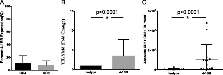
Results: TIL expansion was measured in 19 patient samples. The majority of these TIL were CD4+ T cells and were highly activated. Purified CD8+ T cells produced IFN-γ in response to HLA-matched pancreatic tumor targets. PD-1 blockade and 4-1BB stimulation were demonstrated as effective strategies to improve effective TIL yield, including the production of tumor-reactive pancreatic TIL.

Conclusions: TIL expanded from pancreatic tumors are functional and able to respond to pancreatic tumor associated antigens. PD-1 blockade, 41BB stimulation, and CD8+ T cell enrichment are effective strategies to improve TIL yield and tumor reactivity. These results support the development of adoptive cell therapy strategies using TIL for the treatment of pancreatic cancer.

Keywords: Adoptive cell therapy; Pancreatic cancer; T cells; Tumor infiltrating lymphocytes (TIL).


Figures

Fig. 1
Fig. 1
Characterization of pancreatic TIL after expansion. a Expanded pancreatic TIL were characterized phenotypically by flow cytometry for CD4 and CD8 expression on CD3+ lymphocytes (mean % ± SD). n = 19 (b) TIL were surface stained with CD3, CD4, CD8, and CD25, then further characterized by intracellular staining with Foxp3. Data are reported as mean % ± SD. p < 0.0001 as determined by Wilcoxon matched pairs signed rank test. n = 19. c TIL were additionally surface stained for markers of activation and antigen experience CD69 (i, n = 17), CD27 (ii, n = 11), CD28 (iii, n = 11), GITR (iv, n = 8), 4-1BB (v, n = 18), and CD25 (vi, n = 19) on CD4+ and CD8+ cells within the CD3+ lymphocyte population. Summary data are reported as mean % ± SD
Fig. 2
Fig. 2
PD-1 inhibition yields increased pancreatic TIL. a Expression of PD-1 on TIL in digested pancreatic tumor samples was measured by flow cytometry. Representative data shown in flow plots as well as summary data of mean ± SD (n = 4). b After expansion, TIL were surface stained for CD3, CD4, CD8, and PD-1 and analyzed for PD-1 expression on CD3+ CD4+ or CD3+ CD8+ lymphocytes. Data are reported as mean % ± SD (n = 16). Pancreatic TIL were then propagated from fragments of pancreatic adenocarcinoma specimens in 6000 IU/mL rhIL-2 in the presence of 10 μg/mL α-PD-1 or the isotype control (IgG) antibody. After 2 weeks, total TIL were counted (c), and assessed for viability (d). Fold change was normalized to the isotype control. Column graph displays mean values with positive SD shown. Percentages present the corresponding frequency in the PD-1 or control group (n = 3 patients). Statistical significance was determined by the Wilcoxon signed rank test
Fig. 3
Fig. 3
Addition of 4-1BB agonist improves yield of CD8+ pancreatic TIL. a Pancreatic tumor digests were assessed for 4-1BB expression by flow cytometry. The data represent mean + SD (n = 3). Separately, fragments of pancreatic tumors were initially plated in TIL CM containing 6000 I.U./mL IL-2 and 10 ug/mL anti-4-1BB antibody or IgG4 isotype control. TIL were subsequently cultured according to standard techniques, with the addition of 1 ug/mL anti-4-1BB or isotype. b Viable TIL were counted by trypan blue exclusion after pre-REP expansion and reported as a fold change over isotype control for each patient total (n = 3 patients). Significance was determined by the Wilcoxon signed rank test. c Pancreatic TIL were then analyzed by flow cytometry for CD3, CD4, CD8 expression on live lymphocytes. Absolute numbers were derived from cell counts and percentages based on FACS analysis on a per fragment basis. Statistical significance was determined by the Mann-Whitney Test (n = 3 patients). Data are reported as mean % ± SD
Fig. 4
Fig. 4
Stimulated pancreatic TIL display effector function. a Propagated TIL were stimulated with 25 ng/mL of PMA and 500 nM ionomycin for 18 h, then stained for CD3, CD4, CD8, and CD107a or the corresponding isotype antibodies followed by intracellular staining for IFN-γ and analyzed by flow cytometry. All cells stained with the isotype control were stimulated under the same conditions. Representative FACS plots gated on CD3+ CD8+ TIL are shown. b Scatter plots display the percentage of each indicated population for individual patient data. Each analysis refers to the percentage of IFN-gamma (IFN-g) + and/or CD107a + cells within the CD4 or CD8 gates of the CD3+ lymphocytes. Error bars represent mean % ± SD (n = 12)
Fig. 5
Fig. 5
CD8+ pancreatic TIL maintain effector function after selective expansion. a CD8+ T cells were enriched through magnetic depletion of CD4+ cells from bulk TIL and subject to the rapid expansion protocol (REP). b Representative FACS plots of CD3+ TIL were analyzed for CD4 and CD8 expression before and after the procedure described in a. c CD8-enriched TIL were then stimulated with 25 ng/mL of PMA and 500 nM ionomycin and analyzed for surface CD107a and intracellular IFN-gamma (IFN-g) by flow cytometry
Fig. 6
Fig. 6
Pancreatic TIL produced tumor specific immune response. CD8-enriched post-REP TIL from two patients were cultured with HLA-matched (in black) or mismatched (in gray) pancreatic cancer cell lines at a 1:1 ratio. a The patient was HLA-A typed as A01/24 and matched with the MiaPaca-2 (A24) cell line, and mismatched with the Panc-1 (A02/11) pancreatic tumor cell lines. b The patient was HLA-A typed as A02/24 and matched with the CFPAC-1 (A02/03) and Panc-1 (A02/11) pancreatic tumor cell lines, and mismatched with HPAFII (A01/11) tumor cell line. IFN-gamma levels in supernatants were measured after 48 h and reported as mean ± SEM and statistical significance was determined by the student’s t-test. *, p < 0.05, **, p < 0.01, ***, p < 0.001

References

    1. Hidalgo M. Pancreatic cancer. N Engl J Med. 2010;362(17):1605–1617. doi: 10.1056/NEJMra0901557. - DOI - PubMed
    1. Klein AP. Identifying people at a high risk of developing pancreatic cancer. Nat Rev Cancer. 2013;13(1):66–74. doi: 10.1038/nrc3420. - DOI - PMC - PubMed
    1. Dodson LF, Hawkins WG, Goedegebuure P. Potential targets for pancreatic cancer immunotherapeutics. Immunotherapy. 2011;3(4):517–537. doi: 10.2217/imt.11.10. - DOI - PMC - PubMed
    1. Rosenberg SA, Yang JC, Sherry RM, Kammula US, Hughes MS, Phan GQ, Citrin DE, Restifo NP, Robbins PF, Wunderlich JR, Morton KE, Laurencot CM, Steinberg SM, White DE, Dudley ME. Durable complete responses in heavily pretreated patients with metastatic melanoma using T-cell transfew immunotherapy. Clin Cancer Res. 2011;17(13):4550–4557. doi: 10.1158/1078-0432.CCR-11-0116. - DOI - PMC - PubMed
    1. Rusakiewicz S, Semeraro M, Sarabi M, Desbois M, Locher C, Mendez R, Vimond N, Concha A, Garrido F, Isambert N, Chaigneau L, Le Brun-Ly V, Dubreuil P, Cremer I, Caignard A, Poirier-Colame V, Chaba K, Flament C, Halama N, Jäger D, Eggermont A, Bonvalot S, Commo F, Terrier P, Opolon P, Emile JF, Coindre JM, Kroemer G, Chaput N, Le Cesne A, Blay JY, Zitvogel L. Immune infiltrates are prognostic factors in localized gastrointestinal stromal tumors. Cancer Res. 2013;73(12):3499–3510. doi: 10.1158/0008-5472.CAN-13-0371. - DOI - PubMed

Publication types

MeSH terms