. 2016:2016:7696010.
doi: 10.1155/2016/7696010. Epub 2016 Sep 29.

New Possible Surgical Approaches for the Submammary Adipofascial Flap Based on Its Arterial Supply

Affiliations

New Possible Surgical Approaches for the Submammary Adipofascial Flap Based on Its Arterial Supply

Ehab M Elzawawy et al. Anat Res Int. 2016.

Abstract

Introduction. Submammary adipofascial flap (SMAF) is a valuable option for replacement of the inferior portion of the breast. It is particularly useful for reconstruction of partial mastectomy defects. It is also used to cover breast implants. Most surgeons base this flap cranially on the submammary skin crease, reflecting it back onto the breast. The blood vessels supplying this flap are not well defined, and the harvest of the flap may be compromised due to its uncertain vascularity. The aim of the work was to identify perforator vessels supplying SMAF and define their origin, site, diameter, and length. Materials and Methods. The flap was designed and dissected on both sides in 10 female cadavers. SMAF outline was 10 cm in length and 7 cm in width. The flap was raised carefully from below upwards to identify the perforator vessels supplying it from all directions. These vessels were counted and the following measurements were taken using Vernier caliper: diameter, total length, length inside the flap, and distance below the submammary skin crease. Conclusions. The perforators at the lateral part of the flap took origin from the lateral thoracic, thoracodorsal, and intercostal vessels. They were significantly larger, longer, and of multiple origins than those on the medial part of the flap and this suggests that laterally based flaps will have better blood supply, better viability, and more promising prognosis. Both approaches, medially based and laterally based SMAF, carry a better prognosis and lesser chance for future fat necrosis than the classical cranially based flap.


Figures
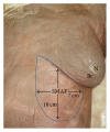
Figure 1
Figure 1
A photograph of left side of female cadaver showing the boundaries of submammary adipofascial flap (SMAF) marked on the skin just below the left breast (Br). Note that the length of the flap is about 10 cm while the width is about 7 cm, and N is the left nipple.
Figure 2
Figure 2
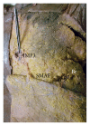
(a) A photograph of the left breast (Br) showing 2 perforators (P1, P2) arising from the superior epigastric artery and passing through rectus abdominis muscle (RA) to supply the upper medial part of SMAF. (b) A close-up photograph or the previous specimen showing P1 and P2. P1 is a long perforator supplying almost the whole width of the upper part of SMAF while P2 divides into 2 branches (A, B).
Figure 3
Figure 3
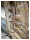
(a) A photograph of anterior abdominal wall showing 7 lateral perforators (P1→P7) on the right side (Rt) and 9 lateral perforators (P1→P9) on the left side (Lt). They supply SMAF. The right nipple (N), the umbilicus (U), and linea alba (LA) are noted. The submammary line is marked by dark line. Xiphoid process is marked by white pin. (b) A close-up photograph of the previous specimen. The perforators arise from the superior epigastric artery (SE) and pass through rectus abdominis muscle (RA) and rectus sheath (RS). (c) A close-up photograph showing the superior epigastric artery (SE) giving medial (M) and lateral (L) perforators. Lateral perforators pass into and supply SMAF.
Figure 4
Figure 4
(a) A photograph of the left breast (Br) showing 3 perforators (IMP3, IMP4, and IMP5) arising from the internal mammary artery and supplying the upper medial part of SMAF. (b) A close-up photograph of the previous specimen. The perforators pass through pectoralis major muscle (PM) to supply the upper medial part of SMAF. IMP4 gives 7 branches (1–7) to SMAF, skin, and the mammary gland.
Figure 5
Figure 5
A photograph of the left breast (Br) showing 2 perforators: IMP3 to the medial part of SMAF and intercostal perforator (ICP) to the lateral part of SMAF.
Figure 6
Figure 6
A photograph of opened anterior chest wall showing the internal mammary artery (A) and vein (V). The artery gives 4 perforators (IMP1, IMP2, IMP3, and IMP5) that pass through the anterior part of the intercostal spaces. It also gives anterior intercostal arteries (IC) passing laterally. IMP4 is missing.
Figure 7
Figure 7
(a) A photograph of the right breast showing the distal part of thoracodorsal artery (TD) giving a perforator (P1) that passes directly to supply the upper lateral part of SMAF. TD also gives P2 that supplies serratus anterior muscle (SA). The right nipple is noted (N). (b) A close-up photograph of the previous specimen showing TD passing through latissimus dorsi muscle (LD). P2 supplies serratus anterior muscle (SA) and passes to the lower lateral part of SMAF. There is a connecting loop between the 2 perforators (arrow).
Figure 8
Figure 8
(a) A photograph of left breast (Br) showing the pectoralis major muscle (PM) cut and reflected to show the axillary artery (AA) and axillary vein (AV) giving the thoracodorsal vessels (TD) that run downwards and give a branch (arrow) to the left breast and the circumflex scapular vessels (arrow head) and terminate as perforator (P) to SMAF. This perforator divides into medial (A) and lateral (B) branches. The intercostobrachial nerve (IB) is noted. (b) A photograph of the right breast (Br) showing the pectoralis major muscle (PM) cut and reflected and the pectoralis minor muscle (Pm) deep to it. The axillary artery (AA) and axillary vein (AV) can be seen. They give the lateral thoracic vessels (LT) which supply the breast itself and give perforator (P1) to SMAF. They also give the thoracodorsal vessels (TD) which give perforator (P2) that communicates through branches (arrows) with P1 and supply SMAF. The distal part of P2 gives 2 capillary perforators, upper (A) and lower (B). Serratus anterior muscle (SA) can be seen. The intercostobrachial nerve (IB) is noted.
Figure 9
Figure 9
(a) A photograph of the left breast (Br) showing the axillary artery (AA) giving the long thoracic artery (LT) which gives perforator (P1) to SMAF and the thoracodorsal artery (TD) which gives muscular branch (M) to latissimus dorsi (LD) and 3 perforators (P2, P3, and P4) to SMAF. A communicating vessel between P1 and P2 is pointed by arrow. P1 gives 2 capillary perforators: upper (A) and lower (B) in SMAF. Thoracoacromial artery (TA) and intercostobrachial nerve (IB) are noted. (b) A photograph of the right breast (Br) showing the axillary artery (AA) giving thoracoacromial artery (TA), long thoracic artery (LT), and thoracodorsal artery (TD). LT gives perforator (P1). TD gives muscular branch (M) to latissimus dorsi (LD) and perforator (P2). P2 divides into medial branch (A) and lateral branch (B). P1 and (A) join and supply SMAF. AA gives a direct perforator (P3) to SMAF. P3 divides into medial branch (C) and lateral branch (D). A communicating vessel between P2 and P3 is pointed by arrow. The intercostobrachial nerve (IB) is noted.
Figure 10
Figure 10
(a) A photograph of the right breast (Br) showing 9 perforators (1→9) supplying SMAF. They arise from the lateral cutaneous branch of the intercostal artery (IC). Right nipple (N) and the xiphoid process (x) are noted. (b) A close-up photograph of the previous specimen showing that each perforator branches into smaller capillary perforators (arrows).
Figure 11
Figure 11
Diagrammatic illustration of the perforators supplying SMAF. LTAP: lateral thoracic artery perforator. TAP: thoracodorsal artery perforator. LICAP: lateral intercostal artery perforator. IMP3: internal mammary perforator 3. IMP4: internal mammary perforator 4. IMP5: internal mammary perforator 5. SEP1: superior epigastric perforator 1. SEP2: superior epigastric perforator 2.

Similar articles

Cited by

References

    1. Jahkola T., Asko-Seljavaara S., Van Smitten K. Immediate breast reconstruction. Scandinavian Journal of Surgery. 2003;92(4):249–256. - PubMed
    1. Dutra A. K., Neto M. S., Garcia É. B., et al. The role of transverse latissimus dorsi musculocutaneous flap immediate breast reconstruction. European Journal of Plastic Surgery. 2009;32(6):293–299. doi: 10.1007/s00238-009-0366-z. - DOI
    1. Healy C., Allen R. J. Sr. The evolution of perforator flap breast reconstruction: twenty years after the first DIEP flap. Journal of Reconstructive Microsurgery. 2014;30(2):121–125. doi: 10.1055/s-0033-1357272. - DOI - PubMed
    1. Freedman G. M., Anderson P. R., Goldstein L. J., et al. Routine mammography is associated with earlier stage disease and greater eligibility for breast conservation in breast carcinoma patients age 40 years and older. Cancer. 2003;98(5):918–925. doi: 10.1002/cncr.11605. - DOI - PubMed
    1. Kijima Y., Yoshinaka H., Funasako Y., et al. Immediate reconstruction using thoracodorsal adipofascial flap after partial mastectomy. Breast. 2009;18(2):126–129. doi: 10.1016/j.breast.2009.02.006. - DOI - PubMed

LinkOut - more resources