

A Review on Recent Developments for Detection of Diabetic Retinopathy
- PMID: 27777811
- PMCID: PMC5061953
- DOI: 10.1155/2016/6838976
A Review on Recent Developments for Detection of Diabetic Retinopathy
Abstract
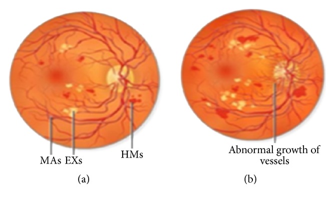
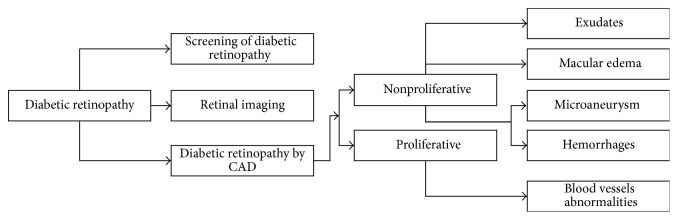
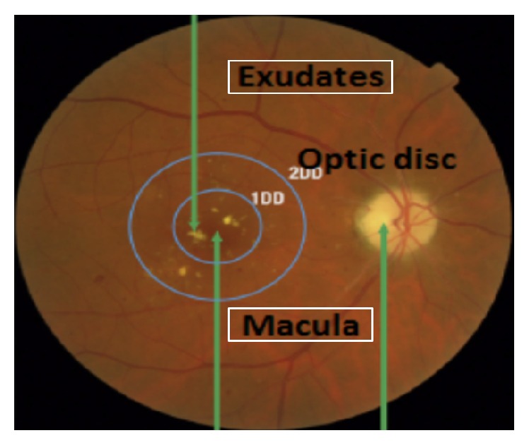
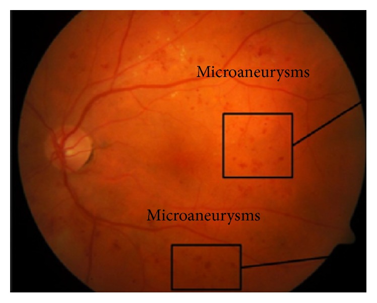
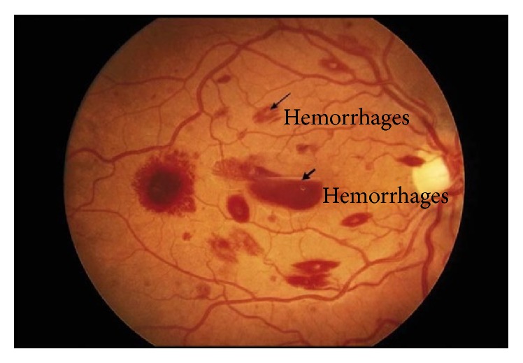
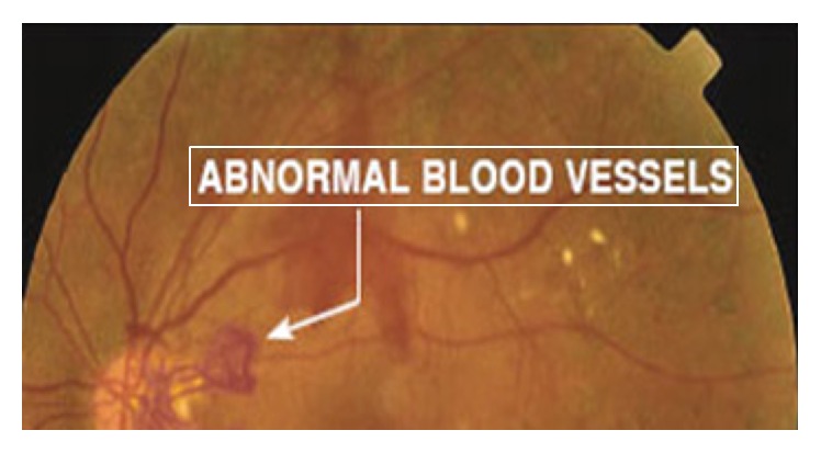
Diabetic retinopathy is caused by the retinal micro vasculature which may be formed as a result of diabetes mellitus. Blindness may appear as a result of unchecked and severe cases of diabetic retinopathy. Manual inspection of fundus images to check morphological changes in microaneurysms, exudates, blood vessels, hemorrhages, and macula is a very time-consuming and tedious work. It can be made easily with the help of computer-aided system and intervariability for the observer. In this paper, several techniques for detecting microaneurysms, hemorrhages, and exudates are discussed for ultimate detection of nonproliferative diabetic retinopathy. Blood vessels detection techniques are also discussed for the diagnosis of proliferative diabetic retinopathy. Furthermore, the paper elaborates a discussion on the experiments accessed by authors for the detection of diabetic retinopathy. This work will be helpful for the researchers and technical persons who want to utilize the ongoing research in this area.
Figures
Similar articles
-
A review on computer-aided recent developments for automatic detection of diabetic retinopathy.J Med Eng Technol. 2019 Feb;43(2):87-99. doi: 10.1080/03091902.2019.1576790. Epub 2019 Jun 14. J Med Eng Technol. 2019. PMID: 31198073 Review.
-
Computer-aided diagnosis of diabetic retinopathy: a review.Comput Biol Med. 2013 Dec;43(12):2136-55. doi: 10.1016/j.compbiomed.2013.10.007. Epub 2013 Oct 14. Comput Biol Med. 2013. PMID: 24290931 Review.
-
Detection of retinal lesions in diabetic retinopathy: comparative evaluation of 7-field digital color photography versus red-free photography.Int Ophthalmol. 2015 Oct;35(5):635-40. doi: 10.1007/s10792-012-9620-7. Epub 2012 Sep 8. Int Ophthalmol. 2015. PMID: 22961609
-
Survey on recent developments in automatic detection of diabetic retinopathy.J Fr Ophtalmol. 2021 Mar;44(3):420-440. doi: 10.1016/j.jfo.2020.08.009. Epub 2021 Jan 30. J Fr Ophtalmol. 2021. PMID: 33526268 Review.
-
Retinal images benchmark for the detection of diabetic retinopathy and clinically significant macular edema (CSME).Biomed Tech (Berl). 2019 May 27;64(3):297-307. doi: 10.1515/bmt-2018-0098. Biomed Tech (Berl). 2019. PMID: 30055096
Cited by
-
Recent developments on computer aided systems for diagnosis of diabetic retinopathy: a review.Multimed Tools Appl. 2023;82(10):14471-14525. doi: 10.1007/s11042-022-13841-9. Epub 2022 Sep 24. Multimed Tools Appl. 2023. PMID: 36185322 Free PMC article.
-
Multi-Label Fundus Image Classification Using Attention Mechanisms and Feature Fusion.Micromachines (Basel). 2022 Jun 15;13(6):947. doi: 10.3390/mi13060947. Micromachines (Basel). 2022. PMID: 35744561 Free PMC article.
-
A Combined Method for Diabetes Mellitus Diagnosis Using Deep Learning, Singular Value Decomposition, and Self-Organizing Map Approaches.Diagnostics (Basel). 2023 May 22;13(10):1821. doi: 10.3390/diagnostics13101821. Diagnostics (Basel). 2023. PMID: 37238305 Free PMC article.
-
Evaluation of Morphological Changes in Retinal Vessels in Type 1 Diabetes Mellitus Patients with the Use of Adaptive Optics.Biomedicines. 2022 Aug 9;10(8):1926. doi: 10.3390/biomedicines10081926. Biomedicines. 2022. PMID: 36009472 Free PMC article.
-
Localization and grading of NPDR lesions using ResNet-18-YOLOv8 model and informative features selection for DR classification based on transfer learning.Heliyon. 2024 May 9;10(10):e30954. doi: 10.1016/j.heliyon.2024.e30954. eCollection 2024 May 30. Heliyon. 2024. PMID: 38779022 Free PMC article.
References
-
- Kauppi T., Kalesnykiene V., Kamarainen J.-K., et al. The DIARETDB1 diabetic retinopathy database and evaluation protocol. Proceedings of the 18th British Machine Vision Conference (BMVC '07); September 2007; pp. 15.1–15.10. - DOI
-
- Kayal D., Banerjee S. A new dynamic thresholding based technique for detection of hard exudates in digital retinal fundus image. Proceedings of the 1st International Conference on Signal Processing and Integrated Networks (SPIN '14); February 2014; pp. 141–144.
-
- Mahendran G., Dhanasekaran R., Narmadha Devi K. N. Identification of exudates for Diabetic Retinopathy based on morphological process and PNN classifier. Proceedings of the 3rd International Conference on Communication and Signal Processing (ICCSP '14); April 2014; Melmaruvathur, India. IEEE; pp. 1117–1121. - DOI
Publication types
LinkOut - more resources
Full Text Sources
Other Literature Sources
