. 2016 Nov;5(4):248-254.
doi: 10.1055/s-0036-1583755. Epub 2016 May 2.

The Etiology and Pathogenesis of Kienböck Disease

Affiliations

The Etiology and Pathogenesis of Kienböck Disease

Gregory Ian Bain et al. J Wrist Surg. 2016 Nov.

Erratum in

Abstract

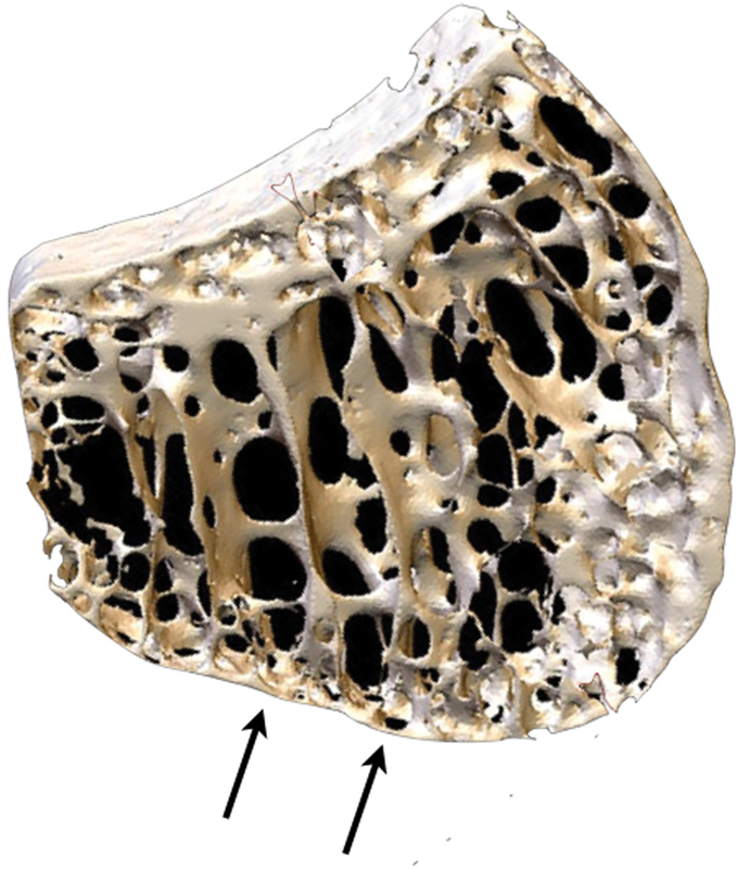
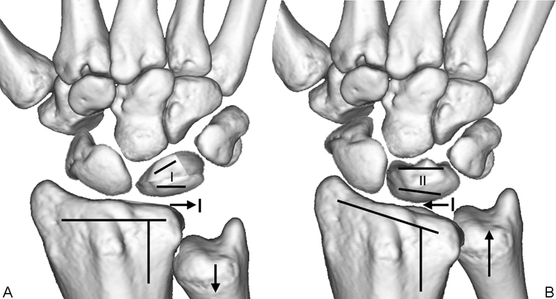
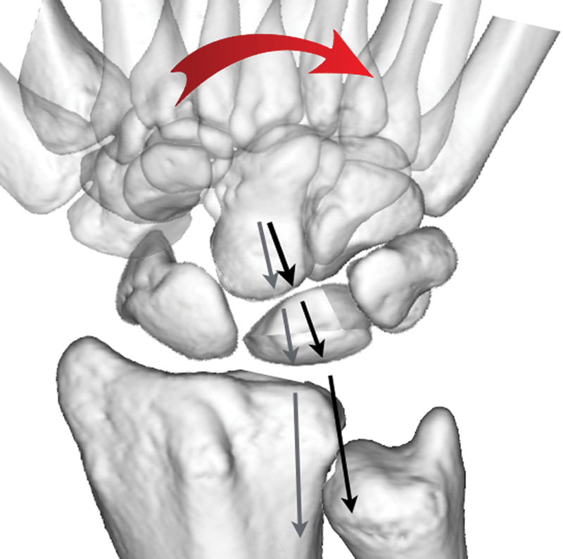
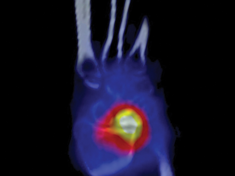
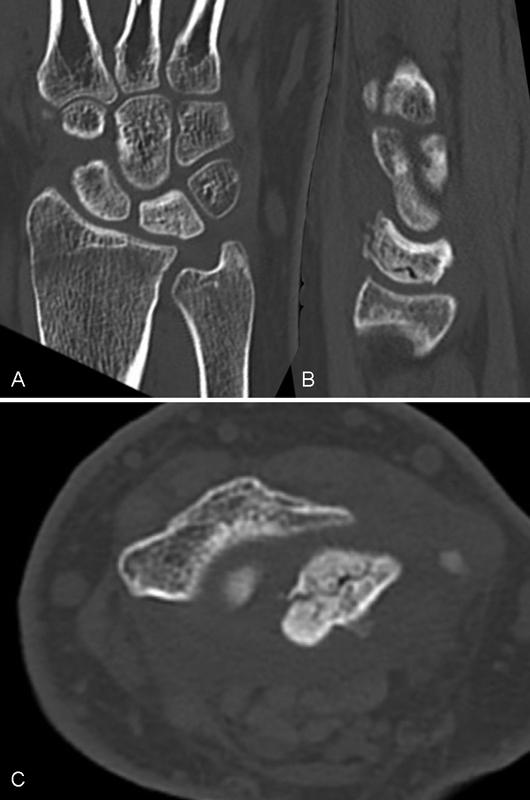
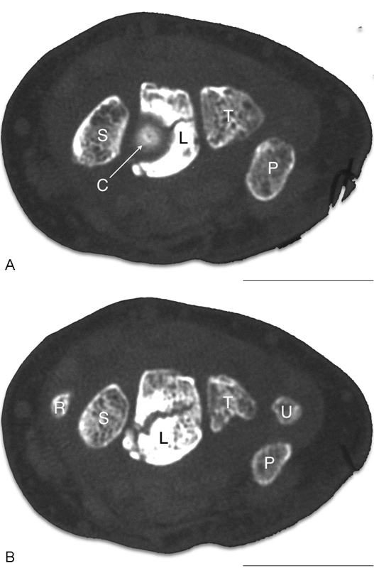
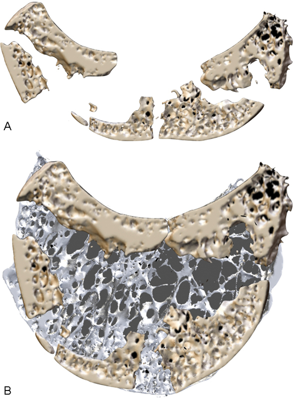
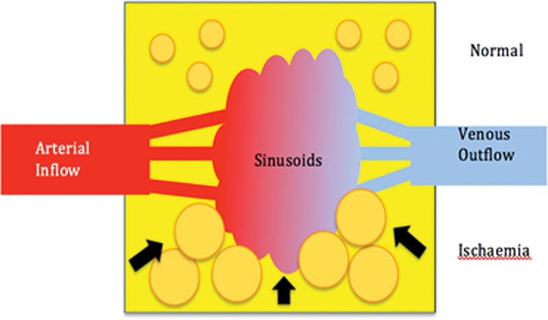
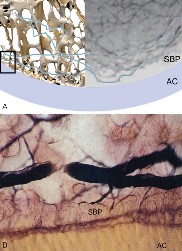
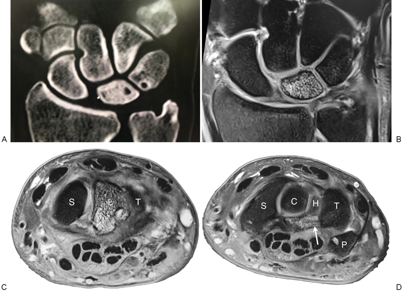
Kienböck disease is a condition that typically occurs in the "at-risk" patient, in the "at-risk" aspect of the proximal condyle of the "at-risk" lunate. In the active male, repetitive loading causes the stress fracture that commences in the single layer proximal subchondral bone plate. The lunate fracture commences at the point the lunate cantilevers over the edge of the distal radius, and then takes on the shape of the radius. We postulate that the stress fracture violates the parallel veins of the venous subarticular plexus-leading to localized venous hypertension and subsequent ischemia and edema of the fatty marrow. The increased osseous compartment pressure further potentiates the venous obstruction, producing avascular necrosis. If the fracture remains localized, it can heal or settle into a stable configuration, so that the wrist remains functional. Fractures of the subchondral bone plate produce irregularity of the lunate articular surfaces and secondary "kissing lesions" of the lunate facet and capitate, and subsequent degeneration. The lunate collapses when the fracture is comminuted, or there is disruption of the spanning trabeculae or a coronal fracture. The secondary effect of the lunate collapse is proximal migration of the capitate between the volar and dorsal fragments, producing collapse of the entire central column. The proximal carpal row is now unstable, and is similar to scapholunate instability, where the capitate migrates between the scaphoid and lunate. The scaphoid is forced into flexion by the trapezium, however, degeneration of the scaphoid and scaphoid facet only occurs in late disease or following failed surgery. In Kienböck disease, the secondary effects of the collapsing lunate are a "compromised" wrist, including: deformity and collapse of the central column, degeneration of the central column (perilunate) articulations, proximal row instability (i.e., between the central and radial columns), and degeneration of the radial column.

Keywords: Kienböck disease; etiology; lunate; pathogenesis.


Conflict of interest statement

Location The Department of Orthopaedic Surgery, Flinders University, Adelaide, South Australia, Australia. Conflict of Interest None.

Figures

Fig. 1
Fig. 1
Microanatomy of the lunate. 3D micro-CT scan of the lunate demonstrating the thin proximal single layer of subchondral bone plate, which was measured to be 0.1-mm thick. There are spanning trabeculae principally on the radial aspect. 3D, three-dimensional; CT, computed tomography. (Image courtesy of Dr. Gregory Bain.)
Fig. 2
Fig. 2
Morphological factors associated with Kienböck disease. (A) The “at-risk” factors increase the load and shear stresses on the radial aspect of the lunate. (B) The “low-risk” factors lead to a wide distribution of the stresses on the lunate.
Fig. 3
Fig. 3
The uncovered type I lunate. The uncovered type I lunate is positioned on the radial edge of the lunate, and with power grip in ulnar deviation loads on the radial edge of the radius.
Fig. 4
Fig. 4
The SPECT scan demonstrates increased uptake within the lunate due to new bone formation and remodeling within the lunate. There is also increased uptake on the radial aspect of the distal radius due to the abnormal loading. SPECT, single-photon emission computed tomography.
Fig. 5
Fig. 5
(A–C) Early case of Kienböck disease. The distribution of the fractures corresponds to the shape of the distal radius. Once the subchondral bone is breached, the friable medullary trabeculae are exposed and comminution and collapse occur.
Fig. 6
Fig. 6
Capitate nutcracker. (A) The capitate is seen at the center of the “target sign” on the radial aspect of the lunate. (B) A coronal fracture propagates across the lunate.
Fig. 7
Fig. 7
Lunate fragmentation. Micro-CT sagittal images of Kienböck lunate: (A) Fragmentation and collapse of the lunate. (B) Reformatted image of the same lunate, superimposed on a normal lunate. Note that most of the cortex is still present but fragmented. However, there is an extensive void, due to resorption of much of the medullary lunate. CT, computed tomography. (Image courtesy of Dr. Gregory Bain.)
Fig. 8
Fig. 8
Compartment syndrome of bone. Diagrammatic representation of the normal vascularization of the lunate (superior half) with the normal fat cell and venous drainage. With ischemia (inferior half), there is interstitial edema and the marrow fat cells become swollen. This leads to tamponade of the sinusoids, thus decreasing the venous outflow. This further increases the intraosseous pressure, reduced arterial inflow, and produces necrosis. (Image courtesy of Dr. Gregory Bain.)
Fig. 9
Fig. 9
Subarticular venous plexus. (A) A composite image of subarticular venous plexus and 3D micro-CT of the subchondral bone plate. The small box on the left of image depicts the area demonstrated in the magnified views show in (B), where the venule within the subarticular plexus seen in profile. The subarticular venous plexus is directly adjacent, making it particularly at risk, even with a stress fracture. The calcified zone of the AC, and SBP are well seen. 3D, three-dimensional; AC, articular cartilage; CT, computed tomography; SBP, subchondral bone plate. (Image courtesy of Crock AO.)
Fig. 10
Fig. 10
Global AVN of the lunate. (A and B) Global sclerosis of the lunate, with large volar perforations of the lunate and triquetrum. We believe this is in part related to the venous hypertension. There is also some localized sclerosis around the venous perforation of the triquetrum. (C and D) Note the changes on the MRI, including the edema of the volar ligaments at the lunotriquetral articulation (white arrow). AVN, avascular necrosis; MRI, magnetic resonance imaging.

References

    1. Kienböck R. Concerning traumatic malacia of the lunate and its consequences: joint degeneration and compression. Fortsch Geb Roentgen. 1910;16:77–103. - PubMed
    1. Müller W. Über die Erweichung und Verdichtung des Os lunatum, eine typische Erkrankung des Handgelenks. Beitr Klin Chir. 1920;119:664.
    1. Ringsted A. Kienböck bei 2 Br ü dern. Acta Chir Scand. 1932;xx:185–196.
    1. Templeman D, Engber W. Kienbock's Disease—Case Report of Familial Occurrence. Iowa Orthop J. 1985;5:107–109.
    1. Gelberman R H, Bauman T D, Menon J, Akeson W H. The vascularity of the lunate bone and Kienböck's disease. J Hand Surg Am. 1980;5(3):272–278. - PubMed