. 2016:2016:4757405.
doi: 10.1155/2016/4757405. Epub 2016 Sep 29.

Crucial Contributions by T Lymphocytes (Effector, Regulatory, and Checkpoint Inhibitor) and Cytokines (TH1, TH2, and TH17) to a Pathological Complete Response Induced by Neoadjuvant Chemotherapy in Women with Breast Cancer

Affiliations

Crucial Contributions by T Lymphocytes (Effector, Regulatory, and Checkpoint Inhibitor) and Cytokines (TH1, TH2, and TH17) to a Pathological Complete Response Induced by Neoadjuvant Chemotherapy in Women with Breast Cancer

Viriya Kaewkangsadan et al. J Immunol Res. 2016.

Abstract

The tumour microenvironment consists of malignant cells, stroma, and immune cells. Prominent tumour-infiltrating lymphocytes (TILs) in breast cancer are associated with a good prognosis and are predictors of a pathological complete response (pCR) with neoadjuvant chemotherapy (NAC). The contribution of different T effector/regulatory cells and cytokines to tumour cell death with NAC requires further characterisation and was investigated in this study. Breast tumours from 33 women with large and locally advanced breast cancers undergoing NAC were immunohistochemically (intratumoural, stromal) assessed for T cell subsets and cytokine expression using labelled antibodies, employing established semiquantitative methods. Prominent levels of TILs and CD4+, CD8+, and CTLA-4+ (stromal) T cells and CD8+ : FOXP3+ ratios were associated with a significant pCR; no association was seen with FOXP3+, CTLA-4+ (intratumoural), and PD-1+ T cells. NAC significantly reduced CD4+, FOXP3+, CTLA-4+ (stromal) (concurrently blood FOXP3+, CTLA-4+ Tregs), and PD-1+ T cells; no reduction was seen with CD8+ and CTLA-4+ (intratumoural) T cells. High post-NAC tumour levels of FOXP3+ T cells, IL-10, and IL-17 were associated with a failed pCR. Our study has characterised further the contribution of T effector/regulatory cells and cytokines to tumour cell death with NAC.


Figures

Figure 1
Figure 1
TILs in the sections of LLABCs, using H&E staining, at 400x magnification; (a) low level of lymphocytic infiltration; (b) high level of lymphocytic infiltration. Low level of TILs defined as ≤60% of tumour nests (Itu: intratumoural) and stromal areas (Str: stromal) infiltrated by lymphocytes. High level of TILs defined as >60% of tumour nests and/or stromal areas infiltrated by lymphocytes.
Figure 2
Figure 2
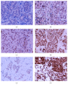
CD4+ (a, b) and CD8+ (c, d) T lymphocytes in the sections of LLABCs, using IHC staining, at 400x magnification. Briefly, heat-mediated antigen retrieval was performed using citrate buffer, pH 6 (20 mins). The sections were then incubated with MAbs to CD4 (Dako, M7310) at a 1 : 80 dilution for 30 mins at RT and MAbs to CD8 (Dako, M7103) at a 1 : 100 dilution for 30 mins at RT. Polymeric HRP-linker antibody conjugate was used as secondary antibody. DAB chromogen was used to visualize the staining. The sections were counterstained with haematoxylin. (a, c) Low level of CD4+ and CD8+T cell infiltration; (b, d) high level of CD4+ and CD8+T cell infiltration. The average number of brown membrane-stained cells, regardless of intensity, in contact with tumour cells or within tumour cell nests (Itu: intratumoural) and in the interstitial stroma (Str: stromal) per HPF was counted.
Figure 3
Figure 3
FOXP3+ (a, b) and CTLA-4+ (c, d) Tregs in the sections of LLABCs, using IHC staining, at 400x magnification. Briefly, heat-mediated antigen retrieval was performed using citrate buffer, pH 6 (20 mins). The sections were then incubated with MAbs to FOXP3 (Abcam, ab20034) at a concentration of 20 μg/mL for 30 mins at RT and MAbs to CTLA-4 (Santa Cruz Bio, sc-376016) at a 1 : 300 dilution for 30 mins at RT. Polymeric HRP-linker antibody conjugate was used as secondary antibody. DAB chromogen was used to visualize the staining. The sections were counterstained with haematoxylin. (a, c) Low level of FOXP3+, CTLA-4+Treg infiltration; (b, d) high level of FOXP3+ and CTLA-4+Treg infiltration. The average number of brown nuclear-stained (FOXP3), membrane-stained (CTLA-4) cells, regardless of intensity, in contact with tumour cells or within tumour cell nests (Itu: intratumoural) and in the interstitial stroma (Str: stromal) per HPF was counted.
Figure 4
Figure 4
PD-1+ T cells in the sections of LLABCs, using IHC staining, at 400x magnification. Briefly, heat-mediated antigen retrieval was performed using citrate buffer, pH 6 (20 mins). The sections were then incubated with MAbs to PD-1 (Abcam, ab52587) at a 1 : 100 dilution for 30 mins at RT. Polymeric HRP-linker antibody conjugate was used as secondary antibody. DAB chromogen was used to visualize the staining. The sections were counterstained with haematoxylin. (a) Low level of PD-1+ T cell infiltration; (b) high level of PD-1+ T cell infiltration. The average number of brown membrane-stained cells, regardless of intensity, in contact with tumour cells or within tumour cell nests (Itu: intratumoural) and in the interstitial stroma (Str: stromal) per HPF was counted.
Figure 5
Figure 5
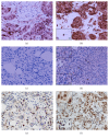
IL-1 (a, b), IL-2 (c, d), and IFN-γ (e, f) expression in the sections of LLABCs, using IHC staining, at 400x magnification. Briefly, heat-mediated antigen retrieval was performed using citrate buffer, pH 6 (20 mins). The sections were then incubated with MAbs to IL-1 (Abcam, ab8320) at a 1 : 150 dilution overnight at 4°C and MAbs to IL-2 (Abcam, ab92381) at a 1 : 500 dilution for 30 mins at RT and polyclonal Abs to IFN-γ (Abcam, ab9657) at a concentration of 4 μg/mL for 30 mins at RT, respectively. Polymeric HRP-linker antibody conjugate was used as secondary antibody. DAB chromogen was used to visualize the staining. The sections were counterstained with haematoxylin. (a, c, e) Low level of expression; (b, d, f) high level of expression. The H score (% of positive cells (brown membrane/cytoplasmic-stained tumour and immune cells) × intensity of staining (1 to 3)) was used to assess the level of expression; low was ≤100 and high was >100. Scoring performed on whole tissue section (>10 HPFs); Tu: tumour and Ly: lymphocyte.
Figure 6
Figure 6
IL-4 (a, b) and IL-10 (c, d) expression in the sections of LLABCs, using IHC staining, at 400x magnification. Briefly, heat-mediated antigen retrieval was performed using citrate buffer, pH 6 (20 mins). The sections were then incubated with polyclonal Abs to IL-4 (Abcam, ab9622) at a concentration of 4 μg/mL for 30 mins at RT and polyclonal Abs to IL-10 (Abcam, ab34843) at a 1 : 400 dilution for 30 mins at RT. Polymeric HRP-linker antibody conjugate was used as secondary antibody. DAB chromogen was used to visualize the staining. The sections were counterstained with haematoxylin. (a, c) Low level of expression; (b, d) high level of expression. The H score (% of positive cells (brown membrane/cytoplasmic-stained tumour and immune cells) × intensity of staining (1 to 3)) was used to assess the level of expression; low was ≤100 and high was >100. Scoring performed on whole tissue section (>10 HPFs); Tu: tumour and Ly: lymphocyte.
Figure 7
Figure 7
IL-17 (a, b), TGF-β (c, d), and PD-L1 (e, f) expression in the sections of LLABCs, using IHC staining, at 400x magnification. Briefly, heat-mediated antigen retrieval was performed using citrate buffer, pH 6 (20 mins). The sections were then incubated with polyclonal Abs to IL-17 (Abcam, ab9565) at a 1 : 100 dilution for 30 mins at RT, MAbs to TGF-β (Abcam, ab64715) at a concentration of 12 μg/mL overnight at 4°C, and polyclonal Abs to PDL1 (Abcam, ab58810) at a concentration of 2.5 μg/mL for 15 mins at RT, respectively. Polymeric HRP-linker antibody conjugate was used as secondary antibody. DAB chromogen was used to visualize the staining. The sections were counterstained with haematoxylin. (a, c, e) Low level of expression; (b, d, f) high level of expression. The H score (% of positive cells (brown membrane/cytoplasmic-stained tumour and immune cells) × intensity of staining (1 to 3)) was used to assess the level of expression; low was ≤100 and high was >100. Scoring performed on whole tissue section (>10 HPFs); Tu: tumour and Ly: lymphocyte.

Similar articles

Cited by

References

    1. Aloysius M., Walker L., Eremin O. Cancer and the immune response. In: Eremin O., Sewell H., editors. Essential Immunology for Surgeons. chapter 4. Oxford, UK: OUP; 2011. pp. 237–302.
    1. Fridman W. H., Pagès F., Sautès-Fridman C., Galon J. The immune contexture in human tumours: impact on clinical outcome. Nature Reviews Cancer. 2012;12(4):298–306. doi: 10.1038/nrc3245. - DOI - PubMed
    1. Galon J., Angell H. K., Bedognetti D., Marincola F. M. The continuum of cancer immunosurveillance: prognostic, predictive, and mechanistic signatures. Immunity. 2013;39(1):11–26. doi: 10.1016/j.immuni.2013.07.008. - DOI - PubMed
    1. Angell H., Galon J. From the immune contexture to the Immunoscore: the role of prognostic and predictive immune markers in cancer. Current Opinion in Immunology. 2013;25(2):261–267. doi: 10.1016/j.coi.2013.03.004. - DOI - PubMed
    1. Teng M. W. L., Ngiow S. F., Ribas A., Smyth M. J. Classifying cancers based on T-cell infiltration and PD-L1. Cancer Research. 2015;75(11):2139–2145. doi: 10.1158/0008-5472.can-15-0255. - DOI - PMC - PubMed

MeSH terms