. 2016 Oct 20;1(17):e87754.
doi: 10.1172/jci.insight.87754.

Improving vascular maturation using noncoding RNAs increases antitumor effect of chemotherapy

Affiliations

Improving vascular maturation using noncoding RNAs increases antitumor effect of chemotherapy

Lingegowda S Mangala et al. JCI Insight. .

Erratum in

  • Improving vascular maturation using noncoding RNAs increases antitumor effect of chemotherapy.
    Mangala LS, Wang H, Jiang D, Wu SY, Somasunderam A, Volk DE, Lokesh GLR, Li X, Pradeep S, Yang X, Haemmerle M, Rodriguez-Aguayo C, Nagaraja AS, Rupaimoole R, Bayraktar E, Bayraktar R, Li L, Tanaka T, Hu W, Ivan C, Gharpure KM, McGuire MH, Thiviyanathan V, Zhang X, Maiti SN, Bulayeva N, Choi HJ, Dorniak PL, Cooper LJ, Rosenblatt KP, Lopez-Berestein G, Gorenstein DG, Sood AK. Mangala LS, et al. JCI Insight. 2021 Apr 8;6(7):e149896. doi: 10.1172/jci.insight.149896. JCI Insight. 2021. PMID: 33793423 Free PMC article. No abstract available.

Expression of concern in

  • Improving vascular maturation using noncoding RNAs increases antitumor effect of chemotherapy.
    Mangala LS, Wang H, Jiang D, Wu SY, Somasunderam A, Volk DE, Lokesh GLR, Li X, Pradeep S, Yang X, Haemmerle M, Rodriguez-Aguayo C, Nagaraja AS, Rupaimoole R, Bayraktar E, Bayraktar R, Li L, Tanaka T, Hu W, Ivan C, Gharpure KM, McGuire MH, Thiviyanathan V, Zhang X, Maiti SN, Bulayeva N, Choi HJ, Dorniak PL, Cooper LJ, Rosenblatt KP, Lopez-Berestein G, Gorenstein DG, Sood AK. Mangala LS, et al. JCI Insight. 2018 Jun 7;3(11):e122387. doi: 10.1172/jci.insight.122387. eCollection 2018 Jun 7. JCI Insight. 2018. PMID: 29889661 Free PMC article. No abstract available.

Abstract

Current antiangiogenesis therapy relies on inhibiting newly developed immature tumor blood vessels and starving tumor cells. This strategy has shown transient and modest efficacy. Here, we report a better approach to target cancer-associated endothelial cells (ECs), reverse permeability and leakiness of tumor blood vessels, and improve delivery of chemotherapeutic agents to the tumor. First, we identified deregulated microRNAs (miRs) from patient-derived cancer-associated ECs. Silencing these miRs led to decreased vascular permeability and increased maturation of blood vessels. Next, we screened a thioaptamer (TA) library to identify TAs selective for tumor-associated ECs. An annexin A2-targeted TA was identified and used for delivery of miR106b-5p and miR30c-5p inhibitors, resulting in vascular maturation and antitumor effects without inducing hypoxia. These findings could have implications for improving vascular-targeted therapy.


Figures
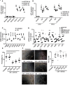
Figure 1
Figure 1. Upregulation of miRs in tumor endothelial cells decreases the expression of tight junction proteins.
(A) Expression of miRs upregulated in patient tumor endothelial cells (ECs) compared with normal ovarian ECs. (BE) ECs were incubated with conditioned medium (CM) from ovarian cancer SKOV3ip1 cells to mimic the tumor microenvironment. (B) Expression of miRs upregulated in tumor-associated ECs compared with untreated ECs. (C) Increased expression of miRs resulted in reduced levels of tight junction proteins. (D) miR silencing increased the expression of tight junction proteins. Fold changes in AD represent the mean of triplicate experiments compared with normal or control or untreated cells. (E) miR silencing decreases the permeability in tumor-associated ECs. miR-inh, miRNA inhibitor. (F) Effect of miR silencing on angiogenesis (tube formation). All experiments were performed 3 times with triplicate samples (n = 3). Values are means ± SEM. *P < 0.05 (1-way ANOVA followed by a Tukey’s multiple comparison post-hoc test). NS, not significant.
Figure 2
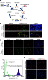
Figure 2. Selection and identification of thioaptamers that bind to human ovarian cancer vascular endothelial cells (ECs).
(A) Cell-SELEX procedure with thioaptamers (TAs). A library of ssDNA TAs was selected, and these TAs were incubated with purified human ovarian cancer ECs, washed, eluted, and amplified for 10 rounds. (B) Representative tumor sections showing binding of Endo28 to human ovarian tumor vasculature. Sections from normal ovary and tumor were incubated with Cy3-labeled Endo28 and anti-CD31 antibody to stain blood vessels. Nuclei are shown in blue, blood vessels in green, and Endo28 in red. Arrows denote colocalization of Endo28 (red) and CD31 (green). (C) Representative tumor sections showing expression of annexin A2 on human ovarian tumor vasculature. Sections from normal ovary and tumor were incubated with anti–annexin A2 and anti-CD31 antibodies. Nuclei are stained blue, annexin A2 green, and blood vessels red. Arrows denote colocalization of CD31 (red) and annexin A2 (green). (D) Annexin A2–positive human microvascular ECs (HMVECs) were detected by flow cytometry. (E) Representative immunofluorescence staining showing that silencing of annexin A2 resulted in reduced binding of Endo28 and Endo31 to HMVECs. Scale bars: 100 μm (C and E).
Figure 3
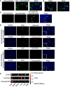
Figure 3. Delivery of CH/Endo28-NPs to tumor vasculature.
Tumors and organs were harvested 6 hours after a single intravenous injection of CH-Endo28-NPs (150 pmol/mouse) into HeyA8 tumor–bearing mice and stained with anti-CD31 antibody to detect blood vessels. (A) Representative tumor sections showing binding of CH/Endo28-NPs to tumor vasculature. Nuclei are shown in blue, blood vessels in green, and CH-Endo28-NPs in red (Cy3). CH/Endo28-NPs, chitosan/Endo28-nanoparticles. (B) Representative tumor sections showing colocalization of CH-Endo28-NPs (red) with blood vessels (green). Arrows in A and B denote colocalization of Endo28 (red) and CD31 (green). (C) Representative tumor sections showing delivery of Alexa 488–conjugated siRNA using CH/Endo28-NPs into tumor cells (blue indicates nuclei, green indicates Alexa 488–conjugated siRNA, and red indicates CH-Endo28-NPs). CH/R4-NPs, chitosan/R4-nanoparticles. Arrows denote colocalization of CH/Endo28 (red) and Alexa 488 siRNA (green).(D) Representative tumor sections showing that annexin A2 silencing reduced delivery of CH/Endo28-NPs to the tumor vasculature. Mice were treated with either CH/control siRNA or CH/mouse annexin A2 (CH/mAnnexin A2) siRNA for 48 hours before a single intravenous injection of CH/R4-NPs or CH/Endo28-NPs. Tumors were harvested 24 hours after NP injection and stained with anti-CD31 antibody to visualize blood vessels. Scale bars: 50 μm. (E) Distribution of CH/Endo28-NPs in tumor cells and organs, examined using IVIS imaging (see Supplemental Methods). Optical imaging of organs and tumors from HeyA8 tumor–bearing mice treated with CH-NPs or CH/R4-NPs or CH/Endo28-NPs shows fluorescence intensity overlaid on white light images of different mouse organs and tumor.
Figure 4
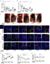
Figure 4. Therapeutic efficacy of CH/Endo28-NPs in a HeyA8 ovarian cancer orthotopic mouse model.
Seven days following tumor cell injection, mice were randomly divided into 6 groups (9–10 mice per group) to receive 1 of the following 6 therapies: (i) CH/Endo28-control miR inhibitor, (ii) CH/Endo28-control miR inhibitor + paclitaxel, (iii) CH/Endo28-miR106b-5p inhibitor, (iv) CH/Endo28-miR106b-5p inhibitor + paclitaxel, (v) CH/Endo28-miR30c-5p inhibitor, or (vi) CH/Endo28-miR30c-5p inhibitor + paclitaxel. Mice were sacrificed when any animals in a control or treatment group became moribund (after 3 to 4 weeks of therapy). (A) Mean tumor weight. (B) Mean number of tumor nodules. (C) Representative images of tumor burden from at least 5 mice per group. (D) Representative tumor sections showing effect of miR silencing on pericyte coverage. Tumor sections were stained with anti-desmin (pericyte coverage marker) and anti-CD31 (for blood vessels) antibodies respectively (red indicates blood vessels, green indicates pericyte coverage, and blue indicates nuclei). (E) Representative tumor sections showing effect of miR silencing on vascular permeability of blood vessels after i.v. injection of FITC-dextran. Tumor sections were stained with anti-CD31 antibody (red indicates blood vessels, green indicates FITC-dextran, and blue indicates nuclei). In D and E, staining was done on 5 sections per group. (F) Determination of extent of hypoxia after staining tumor sections with hypoxia marker anti-CA9 and anti-CD31 antibodies (red indicates blood vessels, green indicates hypoxia, and blue indicates nuclei). (G) Quantification of pericyte coverage and extravasated FITC-dextran. Pericyte coverage was determined by the percentage of vessels with ≥ 50% coverage of desmin in positive cells in 5 random fields of each section at ×200 magnification of each tumor. Quantification of extravasated FITC-dextran was carried out using fluorescence microscopy under the green fluorescent filter using the following scoring system: 0 points, no staining; 1 point, <25%; 2 points, 25%–50%; 3 points, 50%–75%; 4 points, 75%–100% FITC-dextran (using 8–10 sections per tumor at ×200 magnification. (H) Quantification of average number of blood vessels after indicated treatments. To quantify microvessel density, we recorded the number of blood vessels that stained positive for CD31 in 5 random fields of each section at ×200 magnification for each sample. Values are the mean ± SEM. *P < 0.05, **P < 0.01, (1-way ANOVA followed by a Tukey’s multiple comparison post-hoc test). NS, not significant. Scale bars: 100 μm. CH., chitosan; miR-inh, miRNA inhibitor.
Figure 5
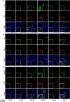
Figure 5. miR silencing increases the expression of tight junction proteins and chemotherapeutic drug delivery.
Seven days following tumor cell injection, mice were randomly divided into 6 groups (9–10 mice per group) to receive 1 of the following 6 therapies: (i) CH/Endo28-control miR inhibitor, (ii) CH/Endo28-control miR inhibitor + paclitaxel, (iii) CH/Endo28-miR106b-5p inhibitor, (iv) CH/Endo28-miR106b-5p inhibitor + paclitaxel, (v) CH/Endo28-miR30c-5p inhibitor, or (vi) CH/Endo28-miR30c-5p inhibitor + paclitaxel. Mice were sacrificed when any animals in a control or treatment group became moribund (after 3 to 4 weeks of therapy). Tumor sections were stained with anti-zo1, -zo2 and –claudin 5 along with -CD31 antibodies. (A) Expression of zo1 (red indicates blood vessels, green indicates zo1). (B) Expression of zo2 (red indicates blood vessels, green indicates zo2). (C) Expression of claudin 5 (red indicates blood vessels, green indicates claudin 5). Staining was done on 5 sections per group (AC). Scale bars: 100 μm. miR-inh, miRNA inhibitor.
Figure 6
Figure 6. Proposed model of effects of miR silencing on restoring tight junction function and reducing vessel permeability.

References

    1. Carmeliet P, Jain RK. Principles and mechanisms of vessel normalization for cancer and other angiogenic diseases. Nat Rev Drug Discov. 2011;10(6):417–427. doi: 10.1038/nrd3455. - DOI - PubMed
    1. Rupaimoole R, et al. Hypoxia-mediated downregulation of miRNA biogenesis promotes tumour progression. Nat Commun. 2014;5:5202. - PMC - PubMed
    1. Bottsford-Miller JN, Coleman RL, Sood AK. Resistance and escape from antiangiogenesis therapy: clinical implications and future strategies. J Clin Oncol. 2012;30(32):4026–4034. doi: 10.1200/JCO.2012.41.9242. - DOI - PMC - PubMed
    1. Sennino B, McDonald DM. Controlling escape from angiogenesis inhibitors. Nat Rev Cancer. 2012;12(10):699–709. doi: 10.1038/nrc3366. - DOI - PMC - PubMed
    1. Zighelboim I, et al. Multicenter phase II trial of topotecan, cisplatin and bevacizumab for recurrent or persistent cervical cancer. Gynecol Oncol. 2013;130(1):64–68. doi: 10.1016/j.ygyno.2013.04.009. - DOI - PMC - PubMed

Publication types