. 2016 Oct 20;1(17):e89402.
doi: 10.1172/jci.insight.89402.

IP3 receptors regulate vascular smooth muscle contractility and hypertension

Affiliations

IP3 receptors regulate vascular smooth muscle contractility and hypertension

Qingsong Lin et al. JCI Insight. .

Abstract

Inositol 1, 4, 5-trisphosphate receptor-mediated (IP3R-mediated) calcium (Ca2+) release has been proposed to play an important role in regulating vascular smooth muscle cell (VSMC) contraction for decades. However, whether and how IP3R regulates blood pressure in vivo remains unclear. To address these questions, we have generated a smooth muscle-specific IP3R triple-knockout (smTKO) mouse model using a tamoxifen-inducible system. In this study, the role of IP3R-mediated Ca2+ release in adult VSMCs on aortic vascular contractility and blood pressure was assessed following tamoxifen induction. We demonstrated that deletion of IP3Rs significantly reduced aortic contractile responses to vasoconstrictors, including phenylephrine, U46619, serotonin, and endothelin 1. Deletion of IP3Rs also dramatically reduced the phosphorylation of MLC20 and MYPT1 induced by U46619. Furthermore, although the basal blood pressure of smTKO mice remained similar to that of wild-type controls, the increase in systolic blood pressure upon chronic infusion of angiotensin II was significantly attenuated in smTKO mice. Taken together, our results demonstrate an important role for IP3R-mediated Ca2+ release in VSMCs in regulating vascular contractility and hypertension.


Figures

Figure 1
Figure 1. Characterization of IP3R deletion in mouse vascular smooth muscle cells in vitro and in vivo.
(A and B) Vascular smooth muscle cells (VSMCs) isolated from noninduced smTKO mice were cultured and treated with 1 μM 4-hydroxytamoxifen (4OHT) for 48 and 72 hours to induce IP3R gene deletion. VSMCs treated with vehicle were used as the control (Ctrl). Administration of 4OHT significantly decreased the mRNA (A) and protein (B) levels of each IP3R subtype compared with the vehicle treatment. n = 3 independent cultures per group. (C and D) mRNA (C) and protein (D) levels of each IP3R subtype in aortas were assessed from control and smTKO mice. n = 3 (with vessels from 3 mice pooled as one sample) per group. (E) Representative curves of Ca2+ release induced by 10 μM ATP in cultured VSMCs treated with vehicle (black) or 4OHT for 72 hours (gray). Cells were incubated with 5 μM fluo-4-AM at 37°C for 30 minutes and imaged with confocal microscopy. Cells were perfused in Ca2+-free solution for 1 to 2 minutes to eliminate Ca2+ entry via membrane ionotropic purinergic receptors prior to the administration of ATP. (F) Averaged Ca2+ signal mass calculated from the time course of Ca2+ release induced by ATP in VSMCs. n = 3 independent tests per group, and at least 20 cells per experiment were imaged. Significance was determined by the 2-tailed, unpaired Student’s t test. *P < 0.05, **P < 0.01, ***P < 0.001.Data represent mean ± SEM.
Figure 2
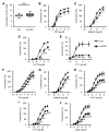
Figure 2. Loss of IP3Rs reduced vascular contractile responses to various vasoconstrictors.
Thoracic aortas and the first-order branch of superior mesenteric arteries were isolated from control (Ctrl) and smTKO mice. The vessels were cut into 2-mm-long segments, and myographic measurements were performed. (A) Reference contraction of aortic rings induced by high potassium (100 mM) in control and smTKO mice. n = 6–11 mice per group. Significance was determined by the 2-tailed, unpaired Student’s t test. In the box-and-whisker plot, the center lines indicate the mean and the upper and lower bounds of the boxes represent the 25th–75th percentiles of the averaged data. (B–E) Tension induced by phenylephrine (PE), U46619, 5-hydroxytryptamine (5-HT), and endothelin 1 (ET1) in control and smTKO aortic rings. (F–J) Tension induced by PE, U46619, 5-HT, ET1, and angiotensin II (AngII) in control and smTKO mesenteric arteries. n = 6 mice per group. For all dose-response curves, data were expressed as a percentage of the peak of K+-induced contraction, and significance was determined by 2-way ANOVA analysis with Bonferroni post-hoc test. *P < 0.05, **P < 0.01, ***P < 0.001 vs. control. Data represent mean ± SEM.
Figure 3
Figure 3. IP3R deficiency repressed U46619-induced myosin light chain 20 and myosin-binding regulatory subunit phosphorylation.
Protein was isolated from control (Ctrl) and smTKO thoracic aortas treated with either vehicle or U46619 (0.1 μM). (A) The phosphorylation of myosin light chain 20 (MLC20) and myosin-binding regulatory subunit (MYPT1) was measured by Western blot. (B) The levels of phosphorylated MLC20 and MYPT1 were normalized to total MLC20 and MYPT1, respectively, and averaged. n = 5 (with vessels from 2 mice pooled as one sample) per group. Significance was determined by 2-way ANOVA analysis with Bonferroni post-hoc test. *P < 0.05, ***P < 0.001 vs. control. #P < 0.05, ##P < 0.01, ###P < 0.001 vs. vehicle. In box-and-whisker plots, center lines indicate the mean and the upper and lower bounds of the boxes represent the 25th–75th percentiles of the averaged data.
Figure 4
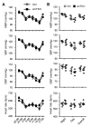
Figure 4. Normal basal blood pressure was observed in smTKO mice.
Blood pressures and heart rate were measured in unrestrained mice using an implantable telemetry system. (A) Twenty-four hours of data are presented at times indicated on a 24-hour scale; data shown at each point represent 3-hour rolling averages of data sampled each minute. The data showed diurnal variations in mean blood pressure (MBP), mean systolic blood pressure (SBP), mean diastolic blood pressure (DBP), and heart rates in both control (Ctrl) and smTKO mice. smTKO mice showed comparable values at all time points compared to control mice. n = 5 mice per group. Significance was determined by 2-way ANOVA analysis with Bonferroni post-hoc test. Data represent mean ± SEM. (B) Mean values for MBP, SBP, DBP, and heart rates were calculated at peak night (20:00–2:00) and day (8:00–14:00) times. n = 5 mice per group. Significance was determined by 2-tailed, unpaired Student’s t test. Data represent mean ± SEM.
Figure 5
Figure 5. Deletion of IP3Rs reduced angiotensin II–induced hypertension.
Angiotensin II (AngII) was infused chronically for 21 consecutive days via a subcutaneously implanted osmotic mini-pump, and blood pressure was measured using the tail-cuff system. (A) Systolic blood pressure (SBP) measured in control (Ctrl) and smTKO mice prior to AngII infusion. n = 6 mice per group. Significance was determined by 2-tailed, unpaired Student’s t test. (B) SBP measured at day –1, day 7, day 14, and day 21 after AngII infusion. n = 6 mice per group. Significance was determined by 2-way ANOVA analysis with Bonferroni post-hoc test. *P < 0.05 vs. control. (C) Representative H&E-stained sections of control and smTKO thoracic aortas before (baseline) and after AngII infusion. Scale bar: 20 μm. (D) Vascular remodeling was accessed by measuring wall thickness and the ratio of medial to luminal area. n = 3–6 mice per group. Significance was determined by 2-way ANOVA analysis with Bonferroni post-hoc test. ##P < 0.01 vs. baseline. Data represent mean ± SEM.

Similar articles

Cited by

References

    1. Conlin PR, Williams GH. Use of calcium channel blockers in hypertension. Adv Intern Med. 1998;43:533–562. - PubMed
    1. Cummings DM, Amadio P, Nelson L, Fitzgerald JM. The role of calcium channel blockers in the treatment of essential hypertension. Arch Intern Med. 1991;151(2):250–259. - PubMed
    1. Haller H. Effective management of hypertension with dihydropyridine calcium channel blocker-based combination therapy in patients at high cardiovascular risk. Int J Clin Pract. 2008;62(5):781–790. doi: 10.1111/j.1742-1241.2008.01713.x. - DOI - PMC - PubMed
    1. De Smedt H, et al. Determination of relative amounts of inositol trisphosphate receptor mRNA isoforms by ratio polymerase chain reaction. J Biol Chem. 1994;269(34):21691–21698. - PubMed
    1. Grayson TH, Haddock RE, Murray TP, Wojcikiewicz RJ, Hill CE. Inositol 1,4,5-trisphosphate receptor subtypes are differentially distributed between smooth muscle and endothelial layers of rat arteries. Cell Calcium. 2004;36(6):447–458. doi: 10.1016/j.ceca.2004.04.005. - DOI - PubMed

Publication types

Substances