. 2016 Oct 20;1(17):e89829.
doi: 10.1172/jci.insight.89829.

The head and neck cancer immune landscape and its immunotherapeutic implications

Affiliations

The head and neck cancer immune landscape and its immunotherapeutic implications

Rajarsi Mandal et al. JCI Insight. .

Abstract

Recent clinical trials have demonstrated a clear survival advantage in advanced head and neck squamous cell carcinoma (HNSCC) patients treated with immune checkpoint blockade. These emerging results reveal that HNSCC is one of the most promising frontiers for immunotherapy research. However, further progress in head and neck immuno-oncology will require a detailed understanding of the immune infiltrative landscape found in these tumors. We leveraged transcriptome data from 280 tumors profiled by The Cancer Genome Atlas (TCGA) to comprehensively characterize the immune landscape of HNSCC in order to develop a rationale for immunotherapeutic strategies in HNSCC and guide clinical investigation. We find that both HPV+ and HPV- HNSCC tumors are among the most highly immune-infiltrated cancer types. Strikingly, HNSCC had the highest median Treg/CD8+ T cell ratio and the highest levels of CD56dim NK cell infiltration, in our pan-cancer analysis of the most immune-infiltrated tumors. CD8+ T cell infiltration and CD56dim NK cell infiltration each correlated with superior survival in HNSCC. Tumors harboring genetic smoking signatures had lower immune infiltration and were associated with poorer survival, suggesting these patients may benefit from immune agonist therapy. These findings illuminate the immune landscape of HPV+ and HPV- HNSCC. Additionally, this landscape provides a potentially novel rationale for investigation of agents targeting modulators of Tregs (e.g., CTLA-4, GITR, ICOS, IDO, and VEGFA) and NK cells (e.g., KIR, TIGIT, and 4-1BB) as adjuncts to anti-PD-1 in the treatment of advanced HNSCC.


Figures

Figure 1
Figure 1. The immune landscape of head and neck squamous cell carcinoma (HNSCC).
(A) Single-sample gene set enrichment analysis for 280 HNSCC tumor samples identifying relative expression of immune cell populations clustered into immune-high and -low phenotypes. (B) Relative cytolytic (CYT) activity, IFN-γ expression, and CD8+/Treg ratios between immune-high and -low tumors clustered by overall immune cell infiltration. (C) Correlations between relative IFN-γ expression and immune infiltration characterized by T cell infiltration score (TIS) and immune cell infiltration score (IIS) in HPV-positive and -negative tumors. (D) Relative TIS, Treg, and CD8+/Treg ratios stratified by head and neck tumor subsite. (E) Relative TIS, IIS, IFN-γ expression, and CYT activity between HNSCC gene expression subtypes. All box-and-whisker plots represent values within the interquartile range (IQR) (boxes) and 1.5 × IQR (whiskers). Outliers are plotted as values > 1.5 × IQR (circles) and > 3 × IQR (stars). All P values for significance (<0.05) represent comparisons via 2-tailed t tests and 1-way ANOVA for continuous comparisons. All r values represent Pearson correlation coefficients. n = 280. APM, antigen-processing machinery.
Figure 2
Figure 2. Immune infiltration and molecular smoking signatures.
(A) T cell infiltration score (TIS), immune cell infiltration score (IIS), and IFN-γ expression scores correlated with the Wellcome Trust Sanger Institute (WTSI) Mutational Smoking Signature Framework in HPV-positive (green) and -negative (blue) tumors. (B) Correlation between nonsynonymous tumor mutational burden and smoking signature quantified by the WTSI Mutational Smoking Signature Framework in HPV-positive (green) and -negative (blue) tumors. All r values represent Pearson correlation coefficients. Two-tailed P values are presented for significance (<0.05). n = 280.
Figure 3
Figure 3. Immune infiltration in HPV-positive and -negative head and neck squamous cell carcinoma (HNSCC).
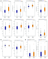
(A) Relative T cell infiltration score (TIS), immune cell infiltration score (IIS), CD8+ T cell infiltration, and Treg infiltration between HPV-negative and -positive squamous cell tumors. (B) Relative expression of immune cell–derived cytotoxic effectors granzyme A, granzyme B, perforin, and their composite effect quantified by their cytolytic (CYT) score between HPV-negative and -positive squamous cell tumors. (C) Relative expression of PD-L1 and CTLA-4 between HPV-negative and -positive squamous cell tumors. (D) Relative median expression of killer cell immunoglobulin-like receptor (KIR) inhibitory genes, KIR2DL1 and KIR2DL3, in HPV-positive and -negative tumors. All box-and-whisker plots represent values within the interquartile range (IQR) (boxes) and 1.5 × IQR (whiskers). All P values for significance (<0.05) represent comparisons via 2-tailed t tests. n = 280.
Figure 4
Figure 4. Pan-cancer immune infiltration analysis.
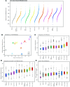
(A) Relative median immune infiltration across the 10 most immune-infiltrated tumors in increasing order including HPV-positive and -negative HNSCC tumors. (B) Correlations between IFN-γ expression scores and median T cell infiltration score (TIS) across the 10 most immune-infiltrated cancer types including HPV-positive and -negative head and neck squamous cell carcinoma (HNSCC) tumors. (C) Relative median Treg infiltration across the 10 most immune-infiltrated tumors in increasing order including HPV-positive and -negative HNSCC tumors. (D) Relative median Treg/CD8+ T cell infiltration ratio across the 10 most immune-infiltrated tumors in increasing order including HPV-positive and -negative HNSCC tumors. (E) Relative median CD56dim NK cell infiltration across the 10 most immune-infiltrated tumors in increasing order including HPV-positive and -negative HNSCC tumors. All box-and-whisker plots represent values within the interquartile range (IQR) (boxes) and 1.5 × IQR (whiskers). Outliers are plotted as values > 1.5 × IQR (circles).
Figure 5
Figure 5. Head and neck squamous cell carcinoma immune infiltration and patient survival.
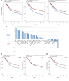
(A) Correlations between expression levels plotted above or below the median of CD8+ T cell infiltration, overall T cell infiltration score (TIS), overall immune cell infiltration score (IIS), and patient cumulative survival. (B) Correlations between patient cumulative survival and immune cell subpopulations and immune effector agents represented by the –log10 P value of the hazard ratio with more positive values indicating better survival and negative values indicating worse survival. (C) Correlation between expression levels plotted above or below the median of Treg infiltration and patient cumulative survival adjusted only for HPV status. Additional correlation between expression levels plotted above or below the median of Treg infiltration and patient cumulative survival adjusted for HPV status, CD8+ TIS, and CD56dim NK cell infiltration, abrogating the survival advantage for Tregs alone. (D) Correlation between high and low expression levels of CD56dim NK cell infiltration and patient cumulative survival adjusted for HPV status. P values for significance (<0.05) calculated using multivariable Cox regression analysis. n = 280.
Figure 6
Figure 6. Head and neck squamous cell carcinoma (HNSCC) immune landscape and implications for immunotherapy.
(A) HNSCC tumors possess the highest median Treg infiltration and the highest Treg/CD8+ T cell infiltration ratio of the 10 most immune-infiltrated tumors, providing a strong rationale to target modulators of Treg function such as CTLA-4, GITR, ICOS, IDO, VEGFA, and others in head and neck cancer immunotherapy. (B) HNSCC tumors possess the highest median CD56dim NK cell infiltration of the 10 most immune-infiltrated tumors providing, an additional rationale to augment NK cell activity in head and neck cancer via modulation of suppressive and agonistic receptors found on NK cells such as KIR, PD-1, TIGIT, 4-1BB, and others. (C) Patients displaying molecular smoking signatures possessed lower immune infiltration of their tumors and displayed worse survival, suggesting these patients may benefit from immune stimulatory therapy such as IL-2, TLR, and STING agonists. Green arrows indicate stimulatory signals and red arrows indicated inhibitory signals.

References

    1. Hamid O, et al. Safety and tumor responses with lambrolizumab (anti-PD-1) in melanoma. N Engl J Med. 2013;369(2):134–144. doi: 10.1056/NEJMoa1305133. - DOI - PMC - PubMed
    1. Hodi FS, et al. Improved survival with ipilimumab in patients with metastatic melanoma. N Engl J Med. 2010;363(8):711–723. doi: 10.1056/NEJMoa1003466. - DOI - PMC - PubMed
    1. Herbst RS, et al. Predictive correlates of response to the anti-PD-L1 antibody MPDL3280A in cancer patients. Nature. 2014;515(7528):563–567. doi: 10.1038/nature14011. - DOI - PMC - PubMed
    1. Tumeh PC, et al. PD-1 blockade induces responses by inhibiting adaptive immune resistance. Nature. 2014;515(7528):568–571. doi: 10.1038/nature13954. - DOI - PMC - PubMed
    1. Tykodi SS. PD-1 as an emerging therapeutic target in renal cell carcinoma: current evidence. Onco Targets Ther. 2014;7:1349–1359. - PMC - PubMed

Publication types