

In vivo Confocal Microscopy Evaluation of Meibomian Gland Dysfunction in Dry Eye Patients with Different Symptoms
- PMID: 27779170
- PMCID: PMC5125342
- DOI: 10.4103/0366-6999.192782
In vivo Confocal Microscopy Evaluation of Meibomian Gland Dysfunction in Dry Eye Patients with Different Symptoms
Abstract
Background: Dry eye patients suffer from all kinds of symptoms. Sometimes, the clinical signs evaluation does not disclose any obvious difference in routine examination; in vivo confocal microscopy (IVCM) is a powerful tool for ocular surface disease. This study aimed to clarify meibomian gland (MG) alterations in dry eye patients with different symptoms and to compare the findings using IVCM.
Methods: A total of sixty patients were recruited, all subjected to Ocular Surface Disease Index (OSDI) and Salisbury Eye Evaluation Questionnaire (SEEQ), and questionnaires for the assessment of dry eye symptoms before clinical sign examinations were given to the patients. Finally, IVCM was applied to observe MG's structure. Statistical analysis was performed using the t-test, Mann-Whitney U-test and Spearman correlation analysis. The differences were statistically significant when P< 0.05.
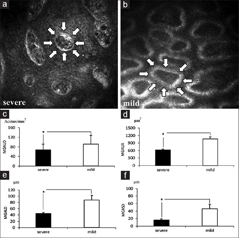
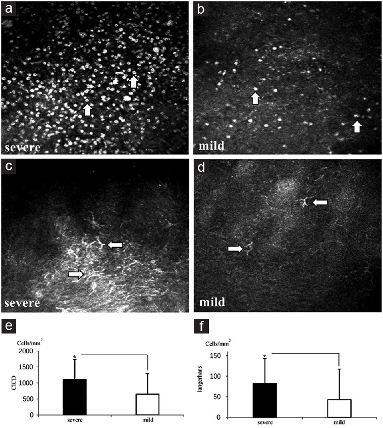
Results: In the severe symptom group, OSDI and SEEQ scores were significantly higher (P< 0.05) compared with the mild symptoms group. All other clinical sign examinations had no statistical difference in the two groups (P> 0.05). However, all the IVCM-observed data showed that patients with severe symptoms had more significant fibrosis in MG (acinar unit area 691.87 ± 182.01 μm2 for the severe, 992.17 ± 170.84 μm2 for the mild; P< 0.05) and severer decrease in the size of MG acinar units than those observed in patients with mild symptoms (MG acinar unit density [MGAUD] 70.08 ± 18.78 glands/mm2, MG acinar unit longest diameter [MGALD] 51.50 ± 15.51 μm, MG acinar unit shortest diameter [MGASD] 20.30 ± 11.85 μm for the severe, MGAUD 89.53 ± 39.88 glands/mm2, MGALD 81.57 ± 21.14 μm, MGASD 42.37 ± 14.55 μm for the mild;P< 0.05). Dry eye symptoms were negatively correlated with MG confocal microscopic parameters and positively correlated with conjunctival inflammatory cells and Langerhans cells (P< 0.05).
Conclusions: IVCM application provides a strong support to differentiate dry eye patients with different symptoms: meibomian gland dysfunction (MGD) plays a pivotal role in dry eye aggravation, and using IVCM to observe MG fibrosis, changes in size and density of MG as well as status of inflammation cells can help not only correctly diagnose the type and severity of dry eye, but also possibly prognosticate in routine eye examination in the occurrence of MGD.
Figures

Similar articles
-
[In vivo confocal microscopy evaluation of meibomian glands in meibomian gland dysfunction patients].Zhonghua Yan Ke Za Zhi. 2016 Sep 11;52(9):649-56. doi: 10.3760/cma.j.issn.0412-4081.2016.09.004. Zhonghua Yan Ke Za Zhi. 2016. PMID: 27647244 Chinese.
-
The efficacy, sensitivity, and specificity of in vivo laser confocal microscopy in the diagnosis of meibomian gland dysfunction.Ophthalmology. 2010 Apr;117(4):665-72. doi: 10.1016/j.ophtha.2009.12.029. Epub 2010 Mar 1. Ophthalmology. 2010. PMID: 20189653 Clinical Trial.
-
Meibomian Gland Alteration in Patients with Primary Chronic Dacryocystitis: An In vivo Confocal Microscopy Study.Curr Eye Res. 2015;40(8):772-9. doi: 10.3109/02713683.2014.959608. Epub 2014 Sep 30. Curr Eye Res. 2015. PMID: 25266812
-
In vivo confocal microscopy in dry eye disease and related conditions.Semin Ophthalmol. 2012 Sep-Nov;27(5-6):138-48. doi: 10.3109/08820538.2012.711416. Semin Ophthalmol. 2012. PMID: 23163268 Free PMC article. Review.
-
Application of In Vivo Confocal Microscopy in Dry Eye Disease.Invest Ophthalmol Vis Sci. 2018 Nov 1;59(14):DES41-DES47. doi: 10.1167/iovs.17-23602. Invest Ophthalmol Vis Sci. 2018. PMID: 30481805 Review.
Cited by
-
Combination therapy with 3% diquafosol tetrasodium ophthalmic solution and sodium hyaluronate: an effective therapy for patients with dry eye after femtosecond laser-assisted in situ keratomileusis.Front Med (Lausanne). 2023 Apr 20;10:1160499. doi: 10.3389/fmed.2023.1160499. eCollection 2023. Front Med (Lausanne). 2023. PMID: 37153094 Free PMC article.
-
Improved Dry Eye Symptoms and Signs of Patients With Meibomian Gland Dysfunction by a Dietary Supplement.Front Med (Lausanne). 2021 Nov 12;8:769132. doi: 10.3389/fmed.2021.769132. eCollection 2021. Front Med (Lausanne). 2021. PMID: 34869485 Free PMC article.
-
The correlation between the microstructure of meibomian glands and ocular Demodex infestation: A retrospective case-control study in a Chinese population.Medicine (Baltimore). 2019 May;98(19):e15595. doi: 10.1097/MD.0000000000015595. Medicine (Baltimore). 2019. PMID: 31083247 Free PMC article.
-
In Vivo Confocal Microscopy in Different Types of Dry Eye and Meibomian Gland Dysfunction.J Clin Med. 2022 Apr 22;11(9):2349. doi: 10.3390/jcm11092349. J Clin Med. 2022. PMID: 35566475 Free PMC article. Review.
-
In Vivo Confocal Microscopy for Automated Detection of Meibomian Gland Dysfunction: A Study Based on Deep Convolutional Neural Networks.J Imaging Inform Med. 2025 Jan 27. doi: 10.1007/s10278-024-01174-y. Online ahead of print. J Imaging Inform Med. 2025. PMID: 39871043
References
-
- The definition and classification of dry eye disease: Report of the definition and classification subcommittee of the International Dry Eye Workshop (2007) Ocul Surf. 2007;5:75–92. - PubMed
-
- Bron AJ, Tiffany JM, Gouveia SM, Yokoi N, Voon LW. Functional aspects of the tear film lipid layer. Exp Eye Res. 2004;78:347–60. doi: 10.1016/j.exer.2003.09.019. - PubMed
-
- Kobayashi A, Yokogawa H, Sugiyama K. In vivo laser confocal microscopy of Bowman's layer of the cornea. Ophthalmology. 2006;113:2203–8. doi: 10.1016/j.ophtha.2006.05.058. - PubMed
-
- Stanek S. Meibomian gland status comparison between active duty personnel and U.S. veterans. Mil Med. 2000;165:591–3. - PubMed
MeSH terms
LinkOut - more resources
Full Text Sources
Other Literature Sources
Medical
