

Form, shape and function: segmented blood flow in the choriocapillaris
- PMID: 27779198
- PMCID: PMC5078844
- DOI: 10.1038/srep35754
Form, shape and function: segmented blood flow in the choriocapillaris
Abstract
The development of fluid transport systems was a key event in the evolution of animals and plants. While within vertebrates branched geometries predominate, the choriocapillaris, which is the microvascular bed that is responsible for the maintenance of the outer retina, has evolved a planar topology. Here we examine the flow and mass transfer properties associated with this unusual geometry. We show that as a result of the form of the choriocapillaris, the blood flow is decomposed into a tessellation of functional vascular segments of various shapes delineated by separation surfaces across which there is no flow, and in the vicinity of which the transport of passive substances is diffusion-limited. The shape of each functional segment is determined by the distribution of arterioles and venules and their respective relative flow rates. We also show that, remarkably, the mass exchange with the outer retina is a function of the shape of each functional segment. In addition to introducing a novel framework in which the structure and function of the metabolite delivery system to the outer retina may be investigated in health and disease, the present work provides a general characterisation of the flow and transfers in multipole Hele-Shaw configurations.
Figures
 . The vicinity of the separation surfaces takes a comparatively prolonged time to be reached by a passive dye filling the flow domain, as shown in (d) (Video S2). This property may be harnessed to visualise the segmentation of the blood flow.
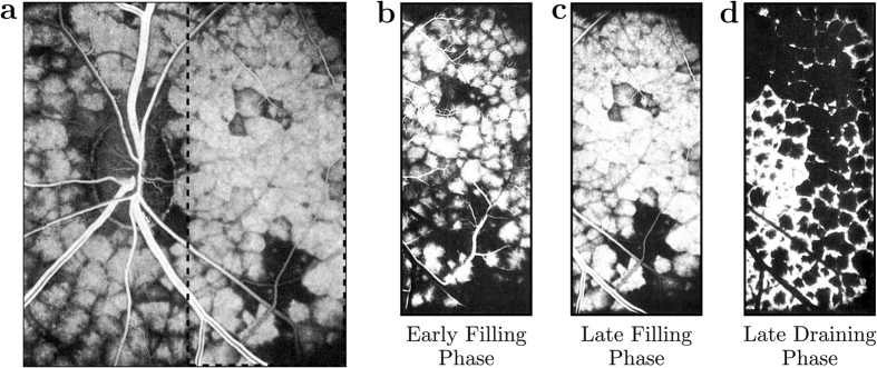
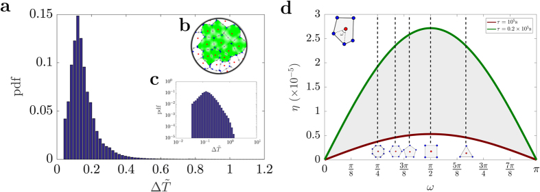
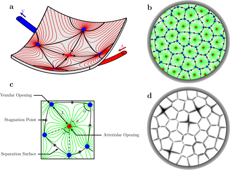
 ) calculated for ten adjacent functional vascular segments of a randomly generated distribution of arteriolar and venular openings (plotted in (b)). In (d), the evolution of the mass extraction η is plotted as a function of the angle ω between an arteriolar opening and two consecutive venular openings feeding and draining the same functional vascular segment (see Methods). Regular polygons associated with certain angles are indicated. The shaded area corresponds to 0.2 × 10⁵ s ≤ τ ≤ 105 s, which concurs with an extraction rate of between 1 and 5% per volume of blood.
References
-
- Hovius J. & Ruysch F. Tractatus de circulari humorum motus in oculi (Langerak, 1702).
-
- Bill A., Sperber G. & Ujiie K. Physiology of the choroidal bed. Int. Ophthalmol. 6, 101–107 (1983). - PubMed
-
- Parver L. M. Temperature modulating action of choroidal blood flow. Eye 5, 181–185 (1991). - PubMed
-
- Labarbera M. Principles of design of fluid transport systems in zoology. Science 249 (1990). - PubMed
Publication types
MeSH terms
LinkOut - more resources
Full Text Sources
Other Literature Sources
Molecular Biology Databases
