

Lumbar Spine Paraspinal Muscle and Intervertebral Disc Height Changes in Astronauts After Long-Duration Spaceflight on the International Space Station
- PMID: 27779600
- PMCID: PMC5588025
- DOI: 10.1097/BRS.0000000000001873
Lumbar Spine Paraspinal Muscle and Intervertebral Disc Height Changes in Astronauts After Long-Duration Spaceflight on the International Space Station
Abstract
Study design: Prospective case series.
Objective: Evaluate lumbar paraspinal muscle (PSM) cross-sectional area and intervertebral disc (IVD) height changes induced by a 6-month space mission on the International Space Station. The long-term objective of this project is to promote spine health and prevent spinal injury during space missions and here on Earth.
Summary of background data: National Aeronautics and Space Administration (NASA) crewmembers have a 4.3 times higher risk of herniated IVDs, compared with the general and military aviator populations. The highest risk occurs during the first year after a mission. Microgravity exposure during long-duration spaceflights results in approximately 5 cm lengthening of body height, spinal pain, and skeletal deconditioning. How the PSMs and IVDs respond during spaceflight is not well described.
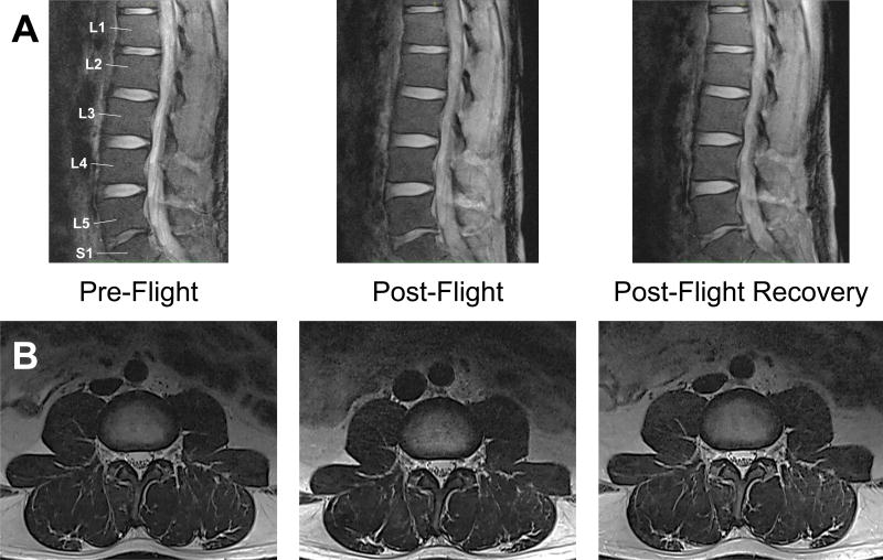
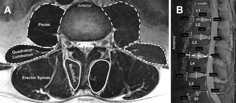
Methods: Six NASA crewmembers were imaged supine with a 3 Tesla magnetic resonance imaging. Imaging was conducted preflight, immediately postflight, and then 33 to 67 days after landing. Functional cross-sectional area (FCSA) measurements of the PSMs were performed at the L3-4 level. FCSA was measured by grayscale thresholding within the posterior lumbar extensors to isolate lean muscle on T2-weighted scans. IVD heights were measured at the anterior, middle, and posterior sections of all lumbar levels. Repeated measures analysis of variance was used to determine significance at P < 0.05, followed by post-hoc testing.
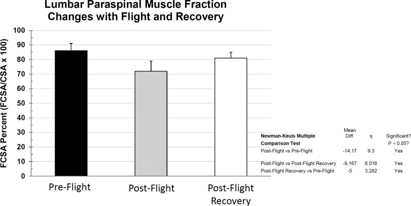
Results: Paraspinal lean muscle mass, as indicated by the FCSA, decreased from 86% of the total PSM cross-sectional area down to 72%, immediately after the mission. Recovery of 68% of the postflight loss occurred during the next 6 weeks, still leaving a significantly lower lean muscle fractional content compared with preflight values. In contrast, lumbar IVD heights were not appreciably different at any time point.
Conclusion: The data reveal lumbar spine PSM atrophy after long-duration spaceflight. Some FCSA recovery was seen with 46 days postflight in a terrestrial environment, but it remained incomplete compared with preflight levels.
Level of evidence: 4.
Figures
Similar articles
-
From the international space station to the clinic: how prolonged unloading may disrupt lumbar spine stability.Spine J. 2018 Jan;18(1):7-14. doi: 10.1016/j.spinee.2017.08.261. Epub 2017 Sep 28. Spine J. 2018. PMID: 28962911 Free PMC article.
-
Negative Effects of Long-duration Spaceflight on Paraspinal Muscle Morphology.Spine (Phila Pa 1976). 2019 Jun 15;44(12):879-886. doi: 10.1097/BRS.0000000000002959. Spine (Phila Pa 1976). 2019. PMID: 30624302
-
Biomechanical changes in the lumbar spine following spaceflight and factors associated with postspaceflight disc herniation.Spine J. 2022 Feb;22(2):197-206. doi: 10.1016/j.spinee.2021.07.021. Epub 2021 Jul 31. Spine J. 2022. PMID: 34343665
-
Neurosurgery and spinal adaptations in spaceflight: A literature review.Clin Neurol Neurosurg. 2021 Aug;207:106755. doi: 10.1016/j.clineuro.2021.106755. Epub 2021 Jun 8. Clin Neurol Neurosurg. 2021. PMID: 34126454 Review.
-
Low Back Pain During and After Spaceflight: A Systematic Review with Meta-Analysis.J Pain Res. 2024 Dec 6;17:4103-4139. doi: 10.2147/JPR.S491060. eCollection 2024. J Pain Res. 2024. PMID: 39660277 Free PMC article. Review.
Cited by
-
Human Health during Space Travel: State-of-the-Art Review.Cells. 2022 Dec 22;12(1):40. doi: 10.3390/cells12010040. Cells. 2022. PMID: 36611835 Free PMC article. Review.
-
Analysis of radiological parameters associated with decreased fractional anisotropy values on diffusion tensor imaging in patients with lumbar spinal stenosis.Eur Spine J. 2019 Jun;28(6):1397-1405. doi: 10.1007/s00586-018-5562-8. Epub 2018 Apr 26. Eur Spine J. 2019. PMID: 29700619
-
Effects of a microgravity SkinSuit on lumbar geometry and kinematics.Eur Spine J. 2023 Mar;32(3):839-847. doi: 10.1007/s00586-022-07454-x. Epub 2023 Jan 16. Eur Spine J. 2023. PMID: 36645514
-
Inhibition of myostatin prevents microgravity-induced loss of skeletal muscle mass and strength.PLoS One. 2020 Apr 21;15(4):e0230818. doi: 10.1371/journal.pone.0230818. eCollection 2020. PLoS One. 2020. PMID: 32315311 Free PMC article.
-
Is a single-level measurement of paraspinal muscle fat infiltration and cross-sectional area representative of the entire lumbar spine?Skeletal Radiol. 2018 Jul;47(7):939-945. doi: 10.1007/s00256-018-2902-z. Epub 2018 Feb 23. Skeletal Radiol. 2018. PMID: 29476224
References
-
- Holt JA, Macias BR, Schneider SM, et al. WISE 2005: Aerobic and resistive countermeasures prevent paraspinal muscle deconditioning during 60-days bed rest in women. J Appl Physiol (1985) 2016 jap 00532 02015. - PubMed
-
- Barr KP, Griggs M, Cadby T. Lumbar stabilization: core concepts and current literature, Part 1. Am. J. Phys. Med. Rehabil. 2005;84(6):473–480. - PubMed
-
- Mooney V, Gulick J, Perlman M, et al. Relationships between myoelectric activity, strength, and MRI of lumbar extensor muscles in back pain patients and normal subjects. J. Spinal Disord. 1997;10(4):348–356. - PubMed
-
- Mannion AF, Weber BR, Dvorak J, Grob D, Muntener M. Fibre type characteristics of the lumbar paraspinal muscles in normal healthy subjects and in patients with low back pain. J. Orthop. Res. 1997;15(6):881–887. - PubMed
MeSH terms
Grants and funding
LinkOut - more resources
Full Text Sources
Other Literature Sources
Medical
Research Materials
Miscellaneous
