Review
. 2017 Mar;150(3):248-264.
doi: 10.1111/imm.12684. Epub 2016 Dec 14.

Deciphering the killer-cell immunoglobulin-like receptor system at super-resolution for natural killer and T-cell biology

Affiliations
Review

Deciphering the killer-cell immunoglobulin-like receptor system at super-resolution for natural killer and T-cell biology

Vivien Béziat et al. Immunology. 2017 Mar.

Abstract

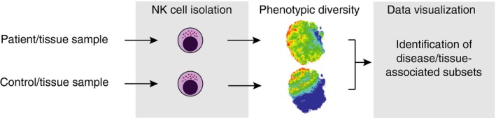
Killer-cell immunoglobulin-like receptors (KIRs) are components of two fundamental biological systems essential for human health and survival. First, they contribute to host immune responses, both innate and adaptive, through their expression by natural killer cells and T cells. Second, KIR play a key role in regulating placentation, and hence reproductive success. Analogous to the diversity of their human leucocyte antigen class I ligands, KIR are extremely polymorphic. In this review, we describe recent developments, fuelled by methodological advances, that are helping to decipher the KIR system in terms of haplotypes, polymorphisms, expression patterns and their ligand interactions. These developments are delivering deeper insight into the relevance of KIR in immune system function, evolution and disease.

Keywords: expression; haplotypes; killer-cell immunoglobulin-like receptors; ligands; natural killer cell; polymorphism.


Figures

Figure 1
Figure 1
Killer‐cell immunoglobulin‐like receptors (KIR) proteins and their ligand interactions. (a) KIR have either two or three immunoglobulin‐like extracellular domains, KIR2D or KIR3D, respectively. They are either activating or inhibitory depending on the structure of their intracellular domain. Inhibitory KIR have long cytoplasmic tails (KIR**L*) that contain immunoreceptor tyrosine‐based inhibitory motifs (ITIM) that transduce inhibitory signals to the natural killer (NK) cell. Activating KIR have short cytoplasmic tails (KIR**S*) with a positively charged amino acid residue in their transmembrane region. The charged residue allows KIR proteins to associate with the TYROBP (DAP12) transmembrane signalling polypeptide, which acts as an activating signal transduction element because it contains an immunoreceptor tyrosine‐based activation motif (ITAM) in its cytoplasmic domain. KIR3DL1 and KIR3DS1, which are encoded by alleles of the same gene, KIR3DL1/S1, thus have opposing functions. KIR differentially bind HLA‐A, ‐B or ‐C allotypes and dimorphisms in the HLA class I α domains are the major determinants for this interaction. The binding motifs are referred to as C1 and C2 in HLA‐C and Bw4 in HLA‐B and HLA‐A. The precise KIR binding motif of HLA‐A*11, which can be recognized by KIR2DS2, KIR2DS4 and KIR3DL2, has not been determined.10, 11 Interactions may also be sensitive to polymorphism outside the HLA and KIR binding motifs and to the presented peptide sequence. The ligands for activating KIR and some inhibitory KIR are presently not well‐defined. OC, open conformers (b) Schematic to show how polymorphism in different parts of the KIR and HLA class I molecules diversifies their interactions. Key residues are KIR position 44 and HLA position 80, which control specificity and KIR position 245 that influences inhibitory signal strength, as discussed in the text.
Figure 2
Figure 2
Structural haplotypes of the KIR gene cluster and recombination mechanisms. Numerous killer‐cell immunoglobulin‐like receptor (KIR) haplotypes with different gene content have been described. These haplotypes have been generated through serial duplications and deletions of chromosomal segments containing KIR genes. The distinction between alleles and genes is, therefore, sometimes blurred; for example KIR2DS3 can be located in two different positions within the KIR locus. (a) The arrangements of genes in 12 common European haplotypes18 are shown. Typically, a person inherits between 14 and 24 KIR genes (between 7 and 12 KIR genes per haplotype). KIR2DP1 and KIR3DP1 are pseudogenes. Two broad haplotypes exist – A (light blue background) and B (pink background), resulting in genotypes that are an ‘AA’, ‘AB’ or ‘BB’. A haplotypes have a single arrangement of seven expressed genes that encode mostly inhibitory KIR, which are diversified by allelic variation. B haplotypes have varied gene arrangements and tend to comprise more activating genes and less allelic diversity. The A haplotype can be divided into two types depending on whether the KIR2DS4 gene is full‐length (KIR2DS4) or carries a frameshift deletion (KIR2DS4 del). (b) Diversity has been generated by homologous recombination, particularly at a recombination hotspot (*) centrally sited within the gene cluster,19 which has shuffled the centromeric (cen) and telomeric (tel) parts of the locus encompassing allelic and gene‐content motifs. (c) Further diversity has been generated through continuing cycles of unequal crossing‐over (non‐allelic homologous recombination), which result in re‐assortment and addition or subtraction of genes in a ‘cut & paste’‐like manner.20, 21 So called fusion genes composed of parts of different KIR genes have been generated by unequal crossover events when the recombination has occurred within genes.21, 22
Figure 3
Figure 3
Major factors that influence natural killer (NK) cell function, expression and killer‐cell immunoglobulin‐like receptors (KIR) ligand binding.
Figure 4
Figure 4
KIR allele counts and KIR3DS1 frequency worldwide plots. (a) Number of alleles reported in the December 2015 release of the IPD KIR database.60 The advent of killer‐cell immunoglobulin‐like receptor (KIR) analysis by next‐generation sequencing is rapidly increasing the number of recognized KIR alleles.68 Blue are genes encoding inhibitory KIR, orange activating KIR and grey are pseudogenes. There are 753 alleles in total. (b) As an example of how KIR gene frequencies can vary significantly across populations, KIR3DS1 gene frequency across the world and (c) Europe are shown (data from The Allele Frequency Net Database56).
Figure 5
Figure 5
From discovery to function. Amino acid residues that have been subject to balancing selection can have dramatic effects on HLA class I recognition. Multiple novel KIR alleles may be discovered during population studies, and molecular analysis identifies the most functionally important. An example is KIR2DL1*022, which was discovered in the southern African KhoeSan population (a) and which differs from its parental allele KIR2DL1*001 by a single nucleotide substitution in codon 44 (b).34 Phylogenetic analyses that included the most closely related KIR from other hominoid species identified that residue 44 has been subject to balancing selection (c).66 Residue 44 occurs at the HLA binding site in the D1 protein domain of the killer‐cell immunoglobulin‐like receptors (KIR) molecule (d) (PDB: 1IM9).70 Substitution of methionine 44 (KIR2DL1*001) for lysine 44 (KIR2DL1*022) switches the specificity of the receptor from HLA‐C2 to HLA‐C1 (e). The methodological pipeline described above links population‐based analyses to functional mapping through sequence/phylogeny analysis and structural biology.
Figure 6
Figure 6
Host genetic variation and functions of HLA class I and killer‐cell immunoglobulin‐like receptors (KIR) in human immunodeficiency virus type 1 (HIV‐1) resistance. HIV‐1 down‐regulates HLA class I expression on the surface of infected CD4+ T cells so as to evade CD8+ T‐cell lysis.86, 87 However, this action exposes the infected cells to recognition and lysis by natural killer (NK) cells through KIRs. The effectiveness of an NK cell response, and consequently the outcome of infection, are linked to the host's KIR genes, their copy number and the expression levels of their ligands. More specifically, resistance to HIV‐1 correlates with; (i) the compound genotype, KIR3DS1HLA‐B Bw4 (with isoleucine at position 80); KIR3DS1 binds HLA‐F open conformers, which can be expressed on HIV‐infected activated CD4+ T cells.88, 89 In this case NK cells expressing KIR3DS1 could also degranulate more potently in response to HIV‐infected Bw4+ CD4+ T cells and suppress viral replication.90 (ii) HLA‐C alleles that confer high cell surface expression91, 92; this may occur because the higher‐expressing HLA alleles result in highly educated NK cells, in addition to more efficient presentation of HIV epitopes to cytotoxic T cells, (iii) high expression KIR3DL1 alleles in Bw4+ individuals; again this could be due to highly educated KIR3DL1+ NK cells with greater activation potential when the ligand is down‐regulated by HIV,58 (iv) Increased copy number of KIR3DS1 alone or KIR3DL1 in the presence of HLA‐B Bw4; probably related to the clonal distribution of KIR, whereby the frequency of NK cells expressing a given KIR correlates linearly with gene copy number, (v) HLA allotypes that form complexes with HIV peptides that bind weakly to inhibitory KIR or strongly to activating KIR. Through any of these mechanisms, NK cells can exert selection pressure on HIV through KIR.63, 93 Conversely, HIV peptides that complex with HLA allotypes and bind inhibitory KIR with high affinity represent NK cell ‘escape’ variants via inhibition of NK cell function.94, 95
Figure 7
Figure 7
KIR genotyping methodology. (a) Representative haplotype‐pairs of two individuals are shown along with the expected results from different typing approaches; (i) presence/absence typing by PCR‐sequence‐specific primers (SSP) or sequence‐specific oligonucleotide (SSO) probes, (ii) copy number typing by quantitative PCR (qPCR),96 multiplex ligation‐dependent probe amplification (MLPA)97 and digital PCR (dPCR),98 (iii) allele and copy number typing by pyrosequencing and next‐generation sequencing (NGS), (iv) imputation infers KIR genotypes from single nucleotide polymorphism (SNP) data. (b) Schemes illustrating different typing approaches. SSP‐specificity relies on single nucleotide differences at the 3′ end of primers to distinguish different KIR genes. Real‐time qPCR combines SSP with fluorescently labelled probes to distinguish KIR genes and a reference gene (always two copies) amplifications in a multiplex reaction. KIR gene copy number is calculated by relative quantification. With NGS typing, oligonucleotide probes are used to capture the KIR genomic region. A bioinformatics pipeline converts sequence data into genotypes. Gene copy number is determined by relative read depth‐ratio of KIR genes compared with a reference gene (always two copies). Allele typing is achieved by filtering reads specific for genes based on alignment to all known reference alleles from KIR coding sequences (e.g. Son of Samtools (SOS)68). In parallel, sequence data can be probed with specific sequence search strings (‘in silico SSO’) to determine which alleles are present (e.g. KIR Filter Fish (KFF)68).
Figure 8
Figure 8
Natural killer (NK) cell strategies in human choriomeningitis virus (HCMV) infection. Viral proteins up‐regulate HLA‐E expression to selectively inhibit NK cells through the NKG2A inhibitory receptor. Concomitantly, the virus down‐regulates classical HLA class I expression to evade CD8+ T cells. NKG2C allows NK cells to detect HLA‐E+ HLA class I‐infected cells. Hence, ‘missing‐self’ (no HLA class I) triggers activation of NK cells already educated/licenced by self‐HLA class I molecules.165, 178 Non‐infected cells are protected from NK cell cytotoxicity by recognition of HLA class I by inhibitory killer‐cell immunoglobulin‐like receptors (KIR), which curbs activation through NKG2C. Activating KIR may directly recognize ‘altered‐self’ ligands that are induced by the virus.15
Figure 9
Figure 9
Natural killer (NK) cell analysis by mass cytometry by time‐of‐flight. By interrogation of multiple markers simultaneously, cell phenotypes can be compared between, for example, patients versus controls or between different tissue sites. The approach allows alterations in lymphocyte composition or abundance to be detected that are linked to killer‐cell immunoglobulin‐like receptors (KIR) genotypes and disease.

References

    1. Battistini L, Borsellino G, Sawicki G, Poccia F, Salvetti M, Ristori G et al Phenotypic and cytokine analysis of human peripheral blood γδ T cells expressing NK cell receptors. J Immunol 1997; 159:3723–30. - PubMed
    1. Young NT, Uhrberg M, Phillips JH, Lanier LL, Parham P. Differential expression of leukocyte receptor complex‐encoded Ig‐like receptors correlates with the transition from effector to memory CTL. J Immunol 2001; 166:3933–41. - PubMed
    1. Patterson S, Chaidos A, Neville DC, Poggi A, Butters TD, Roberts IA et al Human invariant NKT cells display alloreactivity instructed by invariant TCR‐CD1d interaction and killer Ig receptors. J Immunol 2008; 181:3268–76. - PubMed
    1. van Bergen J, Kooy‐Winkelaar EM, van Dongen H, van Gaalen FA, Thompson A, Huizinga TW et al Functional killer Ig‐like receptors on human memory CD4+ T cells specific for cytomegalovirus. J Immunol 2009; 182:4175–82. - PubMed
    1. Anfossi N, Doisne JM, Peyrat MA, Ugolini S, Bonnaud O, Bossy D et al Coordinated expression of Ig‐like inhibitory MHC class I receptors and acquisition of cytotoxic function in human CD8+ T cells. J Immunol 2004; 173:7223–9. - PubMed

Publication types

LinkOut - more resources