. 2016 Dec;157(12):4961-4972.
doi: 10.1210/en.2016-1465. Epub 2016 Oct 25.

Developmental and Thyroid Hormone Regulation of the DNA Methyltransferase 3a Gene in Xenopus Tadpoles

Affiliations

Developmental and Thyroid Hormone Regulation of the DNA Methyltransferase 3a Gene in Xenopus Tadpoles

Yasuhiro Kyono et al. Endocrinology. 2016 Dec.

Abstract

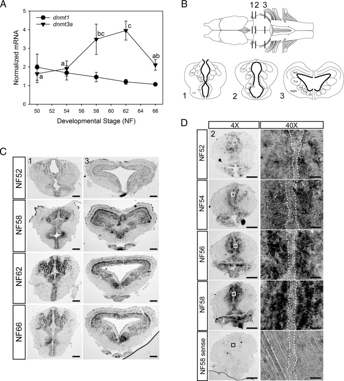
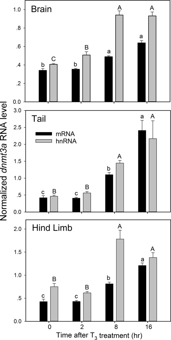
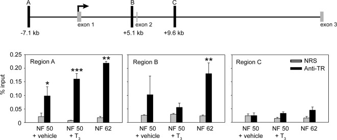
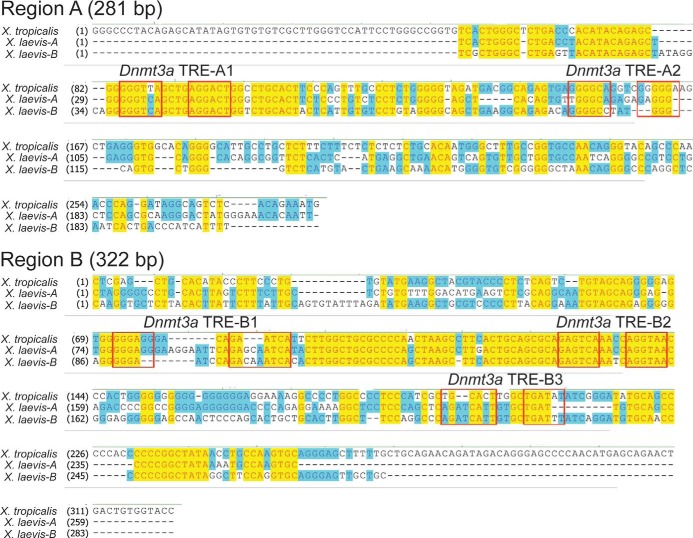
Thyroid hormone is essential for normal development in vertebrates. In amphibians, T3 controls metamorphosis by inducing tissue-specific gene regulation programs. A hallmark of T3 action is the modification of chromatin structure, which underlies changes in gene transcription. We found that mRNA for the de novo DNA methyltransferase (DNMT) dnmt3a, but not dnmt1, increased in the brain of Xenopus tadpoles during metamorphosis in parallel with plasma [T3]. Addition of T3 to the rearing water caused a time-dependent increase in dnmt3a mRNA in tadpole brain, tail, and hind limb. By analyzing data from a genome-wide analysis of T3 receptor (TR) binding in tadpole tail, we identified several putative T3 response elements (TREs) within the dnmt3a locus. Using in vitro DNA binding, transient transfection-reporter, and chromatin immunoprecipitation assays for TRs, we identified two functional TREs at -7.1 kb and +5.1 kb relative to the dnmt3a transcription start site. Sequence alignment showed that these TREs are conserved between two related frog species, X. laevis and X. tropicalis, but not with amniotes. Our previous findings showed that this gene is directly regulated by liganded TRs in mouse brain, and whereas the two mouse TREs are conserved among Eutherian mammals, they are not conserved in Xenopus species. Thus, although T3 regulation of dnmt3a may be an ancient pathway in vertebrates, the genomic sites responsible for hormone regulation may have diverged or arisen by convergent evolution. We hypothesize that direct T3 regulation of dnmt3a may be an important mechanism for modulating global changes in DNA methylation.


Figures

Figure 1.
Figure 1.
Elevation of dnmt3a (but not dnmt1) mRNA levels, and localization of dnmt3a mRNA in Xenopus tadpole brain during metamorphosis. A, Changes in dnmt3a and dnmt1 mRNAs in X. laevis tadpole brain (middle brain region containing the preoptic area and diencephalon) analyzed by RTqPCR. We normalized dnmt3a and dnmt1 mRNAs to the reference gene α-actinin, which did not change across development. Points represent the means ± SEM (n = 5/developmental stage), and means for dnmt3a mRNA with the same letter are not significantly different (F(4,20) = 3.963; P = .017; ANOVA). The dnmt1 mRNA level was not significantly different across metamorphosis, so no statistical symbols are provided for this gene. B, Dorsal view of Xenopus brain (top) and three transverse sections (bottom) at the region of the ventral hypothalamus and thalamic nuclei (region 1) and the optic tectum and tegmentum (regions 2 and 3). The ventricular and subventricular zones are indicated by the bold lines on the transverse sections. P, posterior thalamic nucleus; C, central thalamic nucleus; Lpv, lateral thalamic nucleus, pars posteroventralis; VM, nucleus motorius nervi trigemini; NPv, nucleus of the paraventricular organ; VH, ventral hypothalamic nucleus; LH, lateral hypothalamus; tect, optic tectum; Tor, torus semicircularis; tegm, mesencephalic tegmentum; Ad, dorsal anterior thalamic nucleus; Av, ventral anterior thalamic nucleus; III, nucleus nervi oculomotorii; TP, posterior tuberculum. C, Distribution of dnmt3a mRNA in X. laevis tadpole brain during metamorphosis analyzed by ISHH. Shown are representative photomicrographs of transverse sections at the two brain regions shown in panel B. For region 1, scale bars = 160 μm for NF stages 52 and 59 and 200 μm for NF stages 62 and 66; for region 3, scale bars = 160 μm for NF stage 52 and 200 μm for NF stages 59, 62, and 66. D, The increase in dnmt3a mRNA in tadpole brain during metamorphosis occurs outside of neurogenic zones. We conducted ISHH for dnmt3a mRNA on X. laevis tadpole brain between premetamorphosis and late prometamorphosis. Shown are representative micrographs of transverse sections at the region of the ventral hypothalamus and thalamus (region 2 in panel B). Right panels show a magnified view (40×) of regions indicated by the boxes in the left panels. Areas within dotted lines indicate the VZ/SVZ. Note that dnmt3a mRNA is absent from the VZ/SVZ. Scale bars = 200 μm (4×) and 20 μm (40×). Hybridization with sense probe gave no signal at any developmental stage; only NF stage 58 is shown at the bottom of the figure.
Figure 2.
Figure 2.
Exogenous T3 induces dnmt3a mRNA and hnRNA in early prometamorphic X. tropicalis tadpole brain (middle brain region containing the preoptic area and diencephalon), tail, and hind limb. We treated early prometamorphic (NF stages 53–54) tadpoles with T3 (5 nm) for 0, 2, 8, or 16 hours and measured dnmt3a mRNA and hnRNA by RTqPCR. We normalized dnmt3a mRNA and hnRNA to ef1a mRNA, which did not change with T3 treatment (data not shown). Control reactions without reverse transcriptase were conducted on each RNA sample and produced no amplification. Bars represent the mean ± SEM (n = 4/time point/treatment), and means with the same letter (a, b, c for mRNA; A, B, C for hnRNA) are not significantly different (brain mRNA: F(3,12) = 101.997, P < .0001; brain hnRNA: F(3,12) = 76.795, P < .0001; tail mRNA: F(3,12) = 67.891, P < .0001; tail hnRNA: F(3,12) = 36.235, P < .0001; hind limb mRNA: F(3,12) = 41.84, P < .0001; hind limb hnRNA: F(3,12) = 32.83, P < .0001; ANOVA).
Figure 3.
Figure 3.
Thyroid hormone receptors associate in chromatin with the dnmt3a locus in Xenopus tadpole brain. The schematic diagram (top) shows the location of the putative TR binding sites (black vertical bars; regions A, B, and C) identified by a ChIA-PET experiment conducted on X. tropicalis tail fin chromatin (L. M. Sachs and N. Buisine, unpublished data). Numbers below the bars indicate the distance from the TSS. The TSS was determined by an RNA-PET experiment (30), and the relative positions of the first two coding exons (CDS 1 and 2) are shown. We conducted targeted ChIP assays for TR (bottom) at regions A, B, and C using chromatin isolated from the brains of NF stages 50–52 X. laevis tadpoles treated with vehicle (0.0003% DMSO) or T3 (5 nm) for 48 hours, and chromatin from the brain of tadpoles at metamorphic climax (NF stage 62). Bars represent the mean ± SEM of the ChIP signal expressed as a percentage of input for anti-TR serum or NRS (negative control) (n = 4/group). Asterisks indicate statistically significant differences between the TR ChIP signal and NRS (*, P < .05; **, P < .01; ***, P < .001; Student's independent sample t test).
Figure 4.
Figure 4.
Sequence alignment of regions A and B of X. tropicalis and X. laevis dnmt3a genes. Alignments were done using Clustal W. For X. tropicalis, we used genome build version 4.1, and for X. laevis we used genome build version 5.0 (there are two X. laevis genes owing to its pseudotetraploidy); region C is not shown because we found no evidence for TR association in brain chromatin by ChIP assay (Figure 2). Boxes indicate the locations of predicted DR+4 TRE half-sites numbered in the 5′ to 3′ direction based on the X. tropicalis reference genome. Note that the TRE-A1 and TRE-B2 sequences are conserved between the two frog species.
Figure 5.
Figure 5.
Predicted TREs at the Xenopus dnmt3a locus bind TR-RXR heterodimers in vitro and support T3-dependent transactivation. A, We conducted EMSA using [32P]-labeled oligonucleotide probes corresponding to X. tropicalis dnmt3a TRE-A1 and TRE-B2 sequences (Table 3). We used in vitro synthesized X. laevis TRβ plus RXRα, and in vitro synthesized luciferase as a negative control. The arrow indicates the location of shifted bands due to TR binding to the probes. B, We conducted competitive EMSAs using a [32P]-labeled duplex oligonucleotide probe corresponding to the T3RE-B2 sequence from the X. tropicalis dnmt3a locus (Table 3). Before gel electrophoresis we incubated the probe with in vitro synthesized Xenopus TRβ and RXRα with or without the indicated radioinert oligonucleotides (1 μm). Duplex oligonucleotides with wild-type sequence (wt) or containing single base pair mutations at each of the two half-sites of the indicated TREs (mut; see Table 3) were used as competitors. The arrow indicates the location of shifted bands due to TRβ-RXRα binding to the probe. C, We tested for transactivation activity of DNA fragments corresponding to X. tropicalis dnmt3a regions A (281 bp) and B (322 bp) in transient transfection-reporter assays. After transfection, we treated cells with vehicle (0.1% DMSO) or T3 (30 nm) for 24 hours before conducting dual luciferase assay. These DNA fragments supported T3-dependent transactivation, which was abolished by single base mutations introduced into each of the two half-sites of TRE-A1 and TRE-B2 (Table 3). Bars represent the mean fold change in firefly luciferase activity (normalized to Renilla luciferase activity) relative to vehicle-treated controls (n = 4/treatment). Asterisks indicate statistically significant differences between vehicle and T3-treated cells (P < .001; Student's independent sample t test).

References

    1. Bernal J. Action of thyroid hormone in brain. J Endocrinol Invest. 2002;25(3):268–288. - PubMed
    1. Anderson GW, Schoonover CM, Jones SA. Control of thyroid hormone action in the developing rat brain. Thyroid. 2003;13(11):1039–1056. - PubMed
    1. Gudernatsch JF. Feeding experiments on tadpoles. I. The influence of specific organs given as food on growth and differentiation. A contribution to the knowledge of organs with internal secretion. Wilhelm Roux Arch Entwickl Mech Org. 1912;35(3):457–483.
    1. Denver RJ. Neuroendocrinology of amphibian metamorphosis. In: Shi YB, ed. Current Topics in Developmental Biology: Animal Metamorphosis. Vol 103 Chap 7 San Diego, CA: Elsevier; 2013:195–227. - PubMed
    1. Brown DD, Cai L. Amphibian metamorphosis. Dev Biol. 2007;306(1):20–33. - PMC - PubMed

MeSH terms

Substances