Review
. 2016 Oct 24;26(20):R1039-R1049.
doi: 10.1016/j.cub.2016.09.011.

The Evolving Neural and Genetic Architecture of Vertebrate Olfaction

Affiliations
Review

The Evolving Neural and Genetic Architecture of Vertebrate Olfaction

Daniel M Bear et al. Curr Biol. .

Abstract

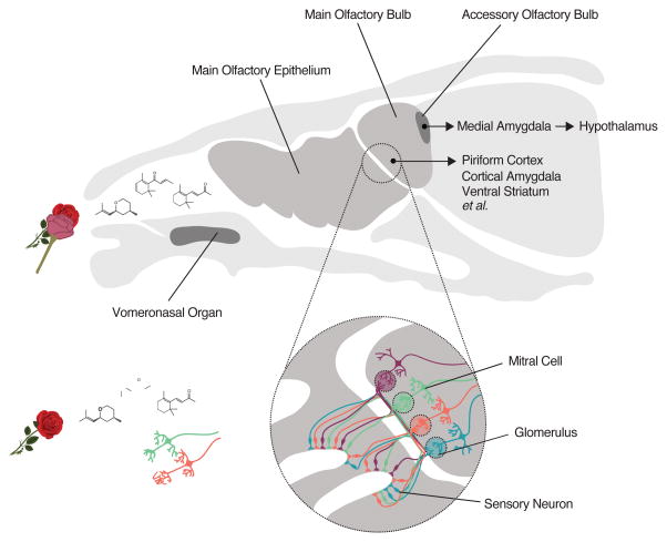
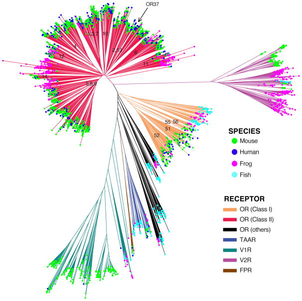
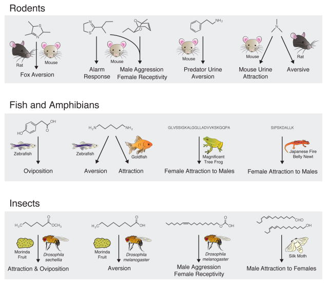
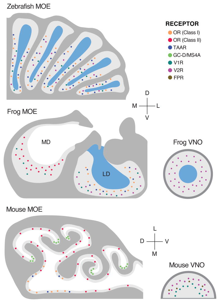
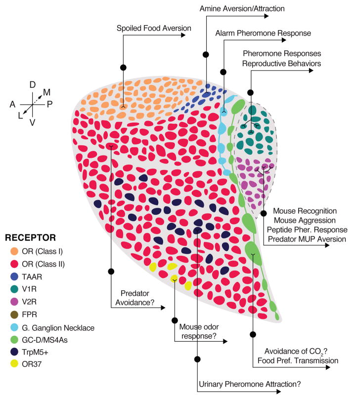
Evolution sculpts the olfactory nervous system in response to the unique sensory challenges facing each species. In vertebrates, dramatic and diverse adaptations to the chemical environment are possible because of the hierarchical structure of the olfactory receptor (OR) gene superfamily: expansion or contraction of OR subfamilies accompanies major changes in habitat and lifestyle; independent selection on OR subfamilies can permit local adaptation or conserved chemical communication; and genetic variation in single OR genes can alter odor percepts and behaviors driven by precise chemical cues. However, this genetic flexibility contrasts with the relatively fixed neural architecture of the vertebrate olfactory system, which requires that new olfactory receptors integrate into segregated and functionally distinct neural pathways. This organization allows evolution to couple critical chemical signals with selectively advantageous responses, but also constrains relationships between olfactory receptors and behavior. The coevolution of the OR repertoire and the olfactory system therefore reveals general principles of how the brain solves specific sensory problems and how it adapts to new ones.


Figures

Figure 1
Figure 1
Organization of the mouse olfactory system (partially adapted from Dulac & Torello, 2003). Odors are blends of molecular compounds inhaled into the nasal cavity, where they interact with olfactory receptor proteins expressed in the main olfactory epithelium (medium gray), the vomeronasal organ (dark gray), or one of several smaller sensory structures not pictured (upper). Each sensory neuron expresses a single olfactory receptor denoted by its color, and neurons expressing the same receptor project to insular structures in the olfactory bulb called glomeruli (lower). Glomeruli corresponding to a given receptor have stereotyped spatial positions across animals. Mitral cells in the olfactory bulb each send a dendrite into one glomerulus (main olfactory bulb) or multiple glomeruli corresponding to the same olfactory receptor (accessory olfactory bulb, not pictured), and project axons to a variety of central brain regions that mediate odor learning and innate odor-driven behaviors. The targets of main and accessory bulb mitral cell projections are largely distinct.
Figure 2
Figure 2
Phylogenetic relationships of the vertebrate GPCR olfactory receptor repertoire. A representative sample of ~2700 GPCR functional OR genes from fish (Danio rerio), frog (Xenopus laevis), mouse (Mus musculus), and human (Homo sapiens) genomes was used to construct a phylogenetic tree (cf. Manzini & Korsching 2011). Notable features are visible, including large expansions of Class II ORs (red) in tetrapods, V2Rs (purple) in amphibians, TAARs (blue) in fishes, and V1Rs (teal) in mammals; significant losses of functional ORs in humans (blue dots) compared to mice (green dots); the monophyly of Class I (orange) and Class II (red) tetrapod ORs; and the subfamily structure of the “classical” ORs, with numeric labels corresponding to the subfamilies defined in Hayden et al. (2010). A Class II OR subfamily (2,13) itself contains a smaller subfamily, the “OR37” receptors, which has atypically expanded and been conserved at the protein sequence level in humans. Note that all mammalian and some frog Class I ORs form a monophyletic clade within the larger set of Class I ORs and other fish OR subfamilies that are neither Class I nor Class II.
Figure 3
Figure 3
A sample of monomolecular odorants that drive innate, species-specific behaviors across vertebrates and insects. Instinctual reproductive, protective, and feeding behaviors are released by a variety of chemical structures, including volatile airborne small molecules in rodents (top row); waterborne acids, bases, and peptides in fishes and amphibians (middle row); and volatile hydrocarbons in insects (bottom row). Chemical structures (receptor responsible for behavior, if known), left to right: top row 2,4,5-trimethythiazoline, 2-sec-butyl-4,5- dihydrothiazole [SBT], 2,3-dehydro-exo-brevicomin [DHB] [76], phenethylamine (Mus musculus TAAR4) [84], trimethylamine (Mus musculus TAAR5) [85]; middle row 4-hydroxyphenylacetic acid (Danio rerio ORA1 [a V1R]) [95], cadaverine (Danio rerio TAAR13c) [58], splendiferin (peptide pheromone for Litoria splendida), sodefrin (peptide pheromone for Cynops pyrrhogaster) [63]; bottom row methylhexanoate (D. sechellia Or22a), octanoic acid [78], cis-11-vaccenyl acetate (D. melanogaster Or67d) [79], (10E,12Z)-hexadeca-10,12-dien-1-al [upper] and (10E,12Z)-hexadeca-10,12-dien-1-ol [lower] (aliases bombykal, receptor Bombyx mori BmOr3, and bombykol, receptor B. mori BmOr1, respectively) [80]. Behavioral responses are sometimes enhanced by combinations of odorants, as with SBT and DHB in mice and bombykal and bombykol in the silk moth. Moreover, responses to the same compound(s) may differ drastically between species or between sexes of the same species.
Figure 4
Figure 4
Peripheral organization of OR family expression across vertebrates. Top: A horizontal section of a quarter of one olfactory rosette from the zebrafish (Danio rerio). Sensory neurons are embedded in a main olfactory epithelium (MOE) with a water-filled lumen. Members of four of the five major GPCR OR families are expressed in different sensory neuron types: TAARs and Class I “classical” ORs [here denoting also fish ORs that belong to neither Class I nor Class II] are expressed in ciliated cells occupying the basal layers of the epithelium; V2Rs in microvillar cells in the middle layer; and at least one V1R in “crypt cells” of the upper layer. Axons of the three cell types project to different, spatially segregated, and stereotyped glomeruli of the zebrafish olfactory bulb. Middle: A coronal section of one half of the main olfactory epithelium and the vomeronasal organ (VNO) of the western clawed frog (Xenopus laevis). An air-filled medial diverticulum (MD) houses neurons expressing Class II “classical” ORs and several V1Rs; a water-filled lateral diverticulum (LD), Class I “classical” ORs, several TAARs, and ancestral V2Rs; the water-filled (VNO), newer members of the expanded V2R family. Bottom: A coronal section of the MOE and VNO of the house mouse (Mus musculus). Receptor family expression is segregated (several exceptions not pictured), with Class I ORs and TAARs expressed in the dorsal MOE, Class II ORs in the ventral MOE (and some in the dorsal MOE, not pictured), V1Rs in the apical VNO, V2Rs in the basal VNO, and FPRs in both VNO layers. Some sensory neurons in the “cul-de-sacs” of the MOE express the receptor guanylate cyclase GC-D and multiple members of a non-GPCR family of four-pass transmembrane chemoreceptor family, Ms4a.
Figure 5
Figure 5
Spatial and molecular organization of projection targets and behavioral responses downstream of distinct mouse olfactory bulb glomeruli. Circumscribed zones of glomeruli are innervated by sensory neurons expressing each of the four largest GPCR OR families or the MS4As. In addition, Class I and Class II OR glomeruli occupy the dorsal-most and more ventral zones, respectively; and projections from Grueneberg Ganglion sensory neurons form their own “necklace” (light blue) anterior to the GC-D/Ms4a “necklace” (green). Dorsal Class II OR glomeruli form a molecularly defined zone, which includes glomeruli necessary for innate aversion of some predator odorants. Within the ventral zone of Class II OR glomeruli, some regions are enriched for the glomeruli of TrpM5- or OR37 subfamily-expressing sensory neurons, which have second-order projections to the “vomeronasal” amygdala and the hypothalamus, respectively.

References

    1. Wyatt TD. Introduction to Chemical Signaling in Vertebrates and Invertebrates. In: Mucignat-Caretta C, editor. Neurobiology of Chemical Communication. Boca Raton (FL): 2014. - PubMed
    1. Meister M. On the dimensionality of odor space. Elife. 2015;4:e07865. - PMC - PubMed
    1. Bargmann CI. Comparative chemosensation from receptors to ecology. Nature. 2006;444:295–301. - PubMed
    1. Hansson BS, Stensmyr MC. Evolution of insect olfaction. Neuron. 2011;72:698–711. - PubMed
    1. Zhang X, Firestein S. The olfactory receptor gene superfamily of the mouse. Nat Neurosci. 2002;5:124–133. - PubMed

Publication types

Substances