. 2016 Dec 2;291(49):25591-25607.
doi: 10.1074/jbc.M116.761296. Epub 2016 Oct 25.

Protein 4.1R Influences Myogenin Protein Stability and Skeletal Muscle Differentiation

Affiliations

Protein 4.1R Influences Myogenin Protein Stability and Skeletal Muscle Differentiation

Shu-Ching Huang et al. J Biol Chem. .

Abstract

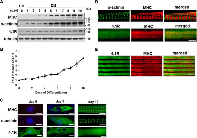
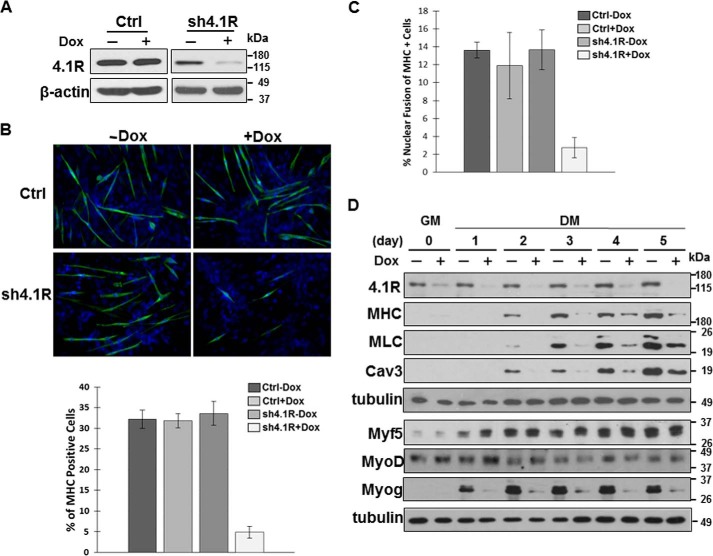
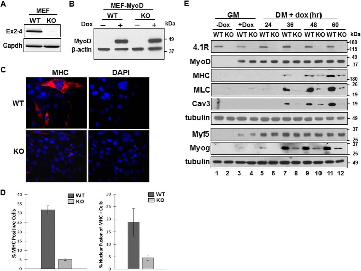
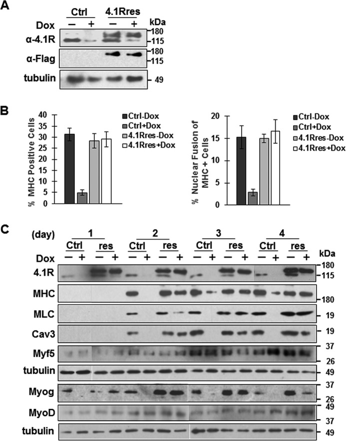
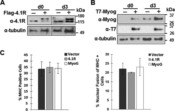
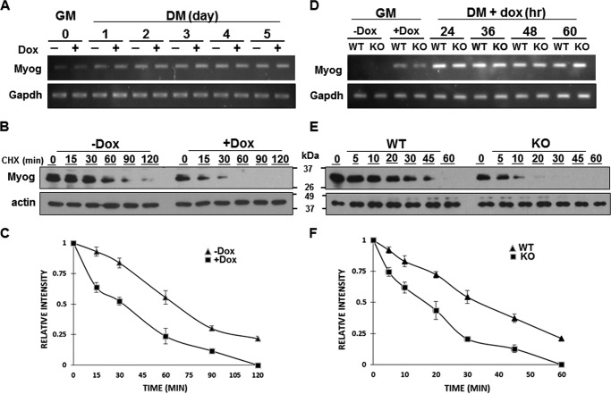
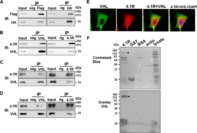
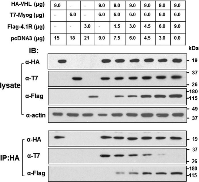
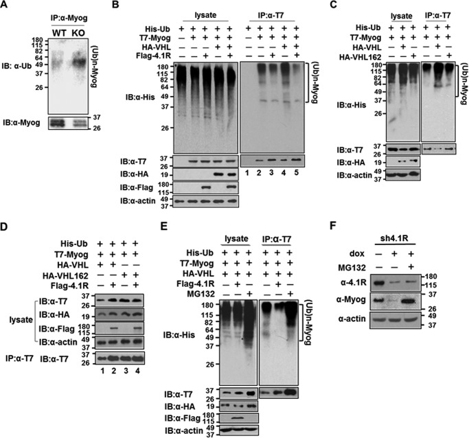
Protein 4.1R (4.1R) isoforms are expressed in both cardiac and skeletal muscle. 4.1R is a component of the contractile apparatus. It is also associated with dystrophin at the sarcolemma in skeletal myofibers. However, the expression and function of 4.1R during myogenesis have not been characterized. We now report that 4.1R expression increases during C2C12 myoblast differentiation into myotubes. Depletion of 4.1R impairs skeletal muscle differentiation and is accompanied by a decrease in the levels of myosin heavy and light chains and caveolin-3. Furthermore, the expression of myogenin at the protein, but not mRNA, level is drastically decreased in 4.1R knockdown myocytes. Similar results were obtained using MyoD-induced differentiation of 4.1R-/- mouse embryonic fibroblast cells. von Hippel-Lindau (VHL) protein is known to destabilize myogenin via the ubiquitin-proteasome pathway. We show that 4.1R associates with VHL and, when overexpressed, reverses myogenin ubiquitination and stability. This suggests that 4.1R may influence myogenesis by preventing VHL-mediated myogenin degradation. Together, our results define a novel biological function for 4.1R in muscle differentiation and provide a molecular mechanism by which 4.1R promotes myogenic differentiation.

Keywords: E3 ubiquitin ligase; myogenesis; myogenin; protein 4.1R; protein degradation; protein stability; ubiquitylation (ubiquitination); von Hippel Lindau.


Figures

FIGURE 1.
FIGURE 1.
Expression and localization of 4.1R, myosin heavy chain, and α-actinin in C2C12 cells during myogenic differentiation. C2C12 cells were cultured in growth medium (GM) and switched to differentiation medium (DM) for a specific number of days as indicated. A, lysates from C2C12 at the indicated days of differentiation were separated by SDS-PAGE and immunoblotted with anti-MHC, anti-α-actinin, anti-4.1R, or anti-β-tubulin antibodies. B, the relative expression of 4.1R in each sample was quantified using densitometry, adjusted for protein loading with β-tubulin, and compared with 4.1R levels in the day 0 sample, which was arbitrarily assigned a value of 1.0. The results represent the means of three separate experiments. C, intracellular localization of 4.1R, MHC, and α-actinin were examined using its respective antibody and revealed with Zeiss microscopy. Day 0 and day 3 cells were also stained with DAPI to visualize the nucleus. Bar, 5 μm. D, double-stained day 10 myotubes with MHC and α-actinin or 4.1R. Bar, 5 μm. E, staining with anti-4.1R (green) and anti-MHC (red) of a longitudinal section of adult mouse skeletal muscle. Bar, 5 μm. Error bars, S.D.
FIGURE 2.
FIGURE 2.
4.1R depletion delays C2C12 myogenic differentiation and reduces myogenic related protein expression. A, validation of 4.1R silencing. C2C12 cells transduced with a control scramble shRNA (Ctrl) or 4.1R shRNA (sh4.1R) in pTRIPZ were stably selected with puromycin and treated with or without doxycycline for 24 h. Cell extracts were analyzed for the efficiency of 4.1R knockdown by immunoblotting with an anti-4.1R Ab. β-Actin served as a loading control. B, 4.1R silencing suppresses myogenic differentiation. Control or sh4.1R stable C2C12 cells were grown for 3 days in differentiation medium in the presence or absence of doxycycline. Differentiation was analyzed by immunofluorescence staining for MHC with an anti-MF20 Ab and FITC-conjugated secondary Ab and revealed with Zeiss microscopy (×40). Nuclei were counterstained with DAPI. The graphs represent the percentage of MHC-positive cells in each treatment obtained from three independent experiments. C, 4.1R silencing reduces myotube formation. sh4.1R or control stable C2C12 cells were grown for 4 days in differentiation medium with or without doxycycline. Cells were stained for MHC and DAPI. Fusion indices were calculated as the percentage of cells containing three or more nuclei within the myosin-positive myocytes. Each experiment was repeated three times, and S.D. values were determined. D, 4.1R silencing decreases myogenic related protein expression. Stable sh4.1R C2C12 lines were grown in growth medium (GM) and switched to differentiation medium (DM) in the presence or absence of doxycycline. Cell lysates were collected at the indicated time course and analyzed for the presence of 4.1R as well as the myogenic differentiation marker MHC, MLC, myofusion marker Cav3, and MRFs (Myf5, MyoD, and myogenin). Each was detected with its respective Ab. Tubulin served as a loading control. Note that to detect several proteins from the same set of experiments, an equal amount of lysate was fractionated in more than one gel and transferred to its respective membrane. When the same membrane was used for more than one antibody, the membrane was stripped between each probing with a limitation of 3–4 strippings per membrane. Each membrane was probed with tubulin as a loading control. Error bars, S.D.
FIGURE 3.
FIGURE 3.
Deletion of 4.1R reduces MyoD-induced differentiation and nuclear fusion in MEFs. A, validation of 4.1R deletion in 4.1R−/− MEF cells. Total RNA isolated from the 4.1R+/+ (WT) and 4.1R−/− (KO) cells were analyzed for the presence of exons 2–4, which contains the translation initiation sites for the 135- and 80-kDa isoforms of 4.1R. GAPDH served as an RT-PCR control. B, WT or KO MEF cells transduced with pTRIPO-3FLAG-MyoD were stably selected with puromycin. Expression of MyoD was analyzed after cells were incubated in differentiation medium in the presence or absence of doxycycline for 24 h. Actin served as a loading control. C, 4.1R deletion reduces myogenic differentiation. 4.1R WT-MyoD and KO-MyoD MEF stable lines were grown in differentiation medium in the presence of doxycycline for 48 h. Differentiation was analyzed by immunofluorescence staining for MHC with an anti-MF20 Ab and Texas Red-conjugated secondary Ab and assessed with Zeiss microscopy (×40). Nuclei were counterstained with DAPI. D, graphical presentation of the effect of 4.1R deletion on differentiation and fusion indices. Left graph, the differentiation indices were calculated as the percentage of MHC-positive cells in each treatment obtained from three independent experiments. Right graph, the fusion indices were calculated as the percentage of cells containing three or more nuclei within the MHC-positive cells. Each experiment was repeated three times, and S.D. values (error bars) were determined. E, 4.1R depletion decreases MHC, MLC, Cav3, and Myog protein expression. WT-MyoD and KO-MyoD MEF cells were grown in growth medium (GM) in the presence or absence of doxycycline for 24 h. Doxycycline-treated MEFs in growth medium were then transited to differentiation medium (DM) in the presence of doxycycline for the indicated periods of time. Cell lysates were collected and immunoblotted for the presence of the indicated protein with its respective Abs. Tubulin served as a loading control.
FIGURE 4.
FIGURE 4.
4.1R isoforms expressed in day 0 undifferentiated and day 8 differentiated C2C12. A, schematic diagram of protein 4.1R and its domains. HP, headpiece; MBD, membrane binding domain; SAB, spectrin-actin binding domain; CTD, C-terminal domain. Previously characterized constitutive sequences are indicated as solid gray boxes, and alternatively spliced cassettes are depicted as shaded boxes. Exon numbers are indicated. B, percentile prevalence of the major 4.1R isoforms identified in day 0 and day 8 differentiated C2C12. 4.1R cDNA libraries constructed from RNA isolated from day 0 and day 8 C2C12 cells were screened with an exon 2′-specific oligonucleotide probe that distinguishes between 135 and 80 kDa. Over 95% of 192 cDNA clones from day 0 and day 8 cells include exon 2′ and encode for 135-kDa forms. The exon compositions of the 135-kDa forms were further PCR-screened using specific oligonucleotides flanking the alternatively spliced exons.
FIGURE 5.
FIGURE 5.
Expression of a 4.1R shRNA-resistant construct rescues the phenotypes associated with 4.1R depletion in C2C12 cells. A control vector (Ctrl) or 4.1R shRNA-resistant construct (4.1Rres) introduced into pTRIPZ-4.1R shRNA C2C12 stable lines was grown in the absence or presence of doxycycline in differentiation medium for the indicated time and analyzed. A, expression of exogenous 4.1R proteins was analyzed after cells were incubated in differentiation medium in the presence or absence of doxycycline for 24 h. Anti-4.1R Ab detects both endogenous and exogenous 4.1R, whereas anti-FLAG Ab detects exogenous 4.1R. Tubulin served as a loading control. B, re-expression of 4.1R reconstitutes myogenic differentiation and myotube formation. Control vector (Ctrl)- or 4.1R shRNA-resistant construct (4.1Rres)-transfected cells were grown for 3 days in differentiation medium in the presence or absence of doxycycline. Differentiation and fusion indices were obtained from three independent experiments. Each experiment was repeated three times, and S.D. values (error bars) were determined. C, re-expression of 4.1R reconstitutes myogenic related protein expression. Control vector- or 4.1R shRNA-resistant construct (res)-transfected cells were grown in differentiation medium in the presence or absence of doxycycline. Cell lysates were collected at the indicated time course and analyzed for the presence of 4.1R as well as MHC, MLC, Cav3, and MRFs (Myf5, MyoD, and myogenin). Each was detected with its respective Ab. Tubulin served as a loading control.
FIGURE 6.
FIGURE 6.
Overexpression 4.1R or myogenin has no effect on normal C2C12 differentiation. FLAG-4.1R or T7-myogenin introduced into C2C12 was grown in differentiation medium for the indicated number of days and analyzed. A, expression of FLAG-4.1R. Anti-4.1R Ab detects both endogenous and exogenous 4.1R. Tubulin served as a loading control. B, expression of T7-myogenin. Anti-myogenin Ab detects both endogenous and exogenous myogenin. Anti-T7 Ab detects exogenously expressed myogenin. Tubulin served as a loading control. C, overexpression of 4.1R or myogenin had no effect on normal C2C12 differentiation. 4.1R- or myogenin-transfected C2C12 cells were grown for 3 days in differentiation medium. Differentiation and fusion indices were obtained from three independent experiments. Each experiment was repeated three times, and S.D. values (error bars) were determined.
FIGURE 7.
FIGURE 7.
Reduced myogenin protein stability in C2C12 and MEF cells lacking 4.1R. A–C, analyses of myogenin in C2C12. A, C2C12 sh4.1R stable lines were cultured in growth medium (GM) or differentiation medium (DM) for the indicated number of days in the presence or absence of doxycycline. Total RNA was isolated and amplified by RT-PCR with limited cycles and examined for the presence of Myog. GAPDH served as a control. B, C2C12 sh4.1R stable cells were cultured in differentiation medium in the absence (−Dox) or presence (+Dox) of doxycycline for 3 days. Cells were exposed to 50 μg/ml CHX for the indicated time, and lysates were probed via Western blotting with an anti-myogenin Ab. Actin served as a loading control. C, Western blots from B were quantified, and the intensity of the myogenin bands was normalized to β-actin and expressed as a percentage of the density measured at time 0. Each experiment was repeated three times, and S.D. values (error bars) were determined. D–F, analyses of myogenin in MEF cells. D, stable 4.1R WT-MyoD and KO-MyoD MEF cells were cultured in the presence or absence of doxycycline-containing growth medium for 24 h. The doxycycline-treated cells were then switched to doxycycline-containing differentiation medium for the indicated times and analyzed for the presence of Myog and GAPDH mRNA by RT-PCR analyses. E, Western blotting analyses for the stability of myogenin. WT-MyoD and KO-MyoD MEF cells cultured in doxycycline-containing differentiation medium for 36 h were treated with CHX over a 60-min time course. The expression of myogenin at the indicated times was analyzed by Western blotting using an anti-myogenin Ab. Actin was used as a loading control. F, the half-life of myogenin was quantified as stated in C. Each experiment was repeated three times, and S.D. values were determined.
FIGURE 8.
FIGURE 8.
4.1R associated and co-localized with VHL. A, C2C12 cells were co-transfected with HA-VHL and FLAG-4.1R and analyzed for the association of VHL and 4.1R in a co-immunoprecipitation assay. Left, cell extracts were precipitated (IP) with a control mouse IgG (mIg) or an anti-FLAG (Flag) Ab. The input extracts and immunoprecipitates were examined by immunoblotting (IB) with anti-HA and anti-FLAG Ab for the presence of HA-VHL and FLAG-4.1R, respectively. Right, extracts were precipitated with a control rabbit IgG (rIg) or an anti-HA Ab and examined with anti-HA and anti-FLAG Abs for the presence of HA-VHL and FLAG-4.1R, respectively. B–D, the association of endogenous VHL and 4.1R was analyzed using day 2 differentiated C2C12 (B), undifferentiated C2C12 (C), and mouse adult skeletal muscle (D) cell lysates in an immunoprecipitation assay. Left, cell extracts were immunoprecipitated with a control mouse IgG or an anti-VHL Ab and examined with anti-4.1R and anti-VHL Abs for the presence of 4.1R and VHL, respectively. Right, extracts were precipitated with rabbit Ig or an anti-4.1R Ab and examined with anti-4.1R and anti-VHL Abs for the presence of 4.1R and VHL, respectively. E, C2C12 cells were immunostained with anti-VHL (green) and anti-4.1R (red) and revealed with Zeiss microscopy (×100). DAPI stains DNA (blue). F, blot overlay assays of 4.1R and VHL. Top, 4.1R, GST, BSA, actin, and BL21 lysates are shown by Coomassie Blue staining. The GST-4.1R fusion protein is highly insoluble in the bacteria expression system. Attempts to extract the fusion protein also co-extracted several host proteins. The arrow indicates the 4.1R cleaved from GST-4.1R with PreScission protease. The smaller bands are the contaminated host proteins. Last lane, 50 μg of BL21 lysates was loaded and served as a negative control for co-purified host proteins from 4.1R preparations. Bottom, a duplicate gel with the same amount of proteins as in the Coomassie Blue-stained gel was separated by SDS-PAGE, overlaid with purified His-VHL, and followed by Western blotting with an anti-His antibody. Arrow, 4.1R cleaved from GST-4.1R with PreScission protease.
FIGURE 9.
FIGURE 9.
4.1R competitively interrupts the association of VHL and myogenin. C2C12 cells grown in 10-cm dishes were co-transfected with various combinations of HA-VHL, T7-myogenin, FLAG-4.1R, and empty vector pcDNA3, with a total of 24 μg of DNA, as indicated. Cells were lysed in IP buffer 48 h post-transfection and immunoprecipitated (IP) with an anti-HA antibody. lysate, equal amounts of lysates were analyzed via Western blotting (IB) for the exogenously expressed HA-VHL, T7-Myog, and FLAG-4.1R. IP:HA, anti-HA immunoprecipitates were equally divided and analyzed for HA-VHL and its associated T7-Myog and FLAG-4.1R with their respective antibodies.
FIGURE 10.
FIGURE 10.
4.1R inhibits VHL-mediated ubiquitination and myogenin degradation via the ubiquitin-proteasome pathway. A, 4.1R knockdown increases polyubiquitinated myogenin. WT-MyoD and KO-MyoD MEF cells were cultured in differentiation medium in the presence of doxycycline for 48 h and analyzed for myogenin levels and its status of ubiquitination. Top, ubiquitination of myogenin was analyzed by immunoprecipitation (IP) of cell lysates with an anti-myogenin Ab and immunoblotting (IB) with an anti-Ub antibody. (Ub)n-MyoG, ubiquitinated myogenin. Bottom, the amount of immunoprecipitated myogenin was detected using an anti-myogenin Ab. B, decrease in polyubiquitinated myogenin by 4.1R overexpression. HEK-293 cells were transfected with combinations of His-Ub, T7-Myog, HA-VHL, and FLAG-4.1R as indicated. Left panel, lysates fractionated on an SDS-polyacrylamide gel were blotted with an anti-His antibody. The presence of the exogenously expressed proteins in cell lysates was confirmed by immunoblotting with an anti-T7 Ab for myogenin, anti-HA for VHL, and anti-FLAG for 4.1R. Actin served as a loading control. Right, extracts were immunoprecipitated with an anti-T7 antibody and immunoblotted with an anti-His Ab. The presence of T7-myogenin was confirmed by immunoblotting using an anti-T7 Ab. C, VHL increases the ubiquitination of myogenin. HEK-293 cells were transfected with His-Ub and T7-Myog and in combination with VHL or VHL C162F. Left, extracts were blotted with an anti-His Ab for the presence of ubiquitinated proteins, anti-T7 Ab for T7-Myog, and anti-HA Ab for VHL or VHL C162F. Right, anti-T7 immunoprecipitates were blotted with an anti-His Ab for the ubiquitinated T7-myogenin and anti-T7 Ab for the precipitated T7-Myog. D, 4.1R reverses the ubiquitination and quantity of myogenin in a VHL-specific manner. HEK-293 cells were transfected with His-Ub, T7-Myog, and either HA-VHL or HA-VHL C162F in the presence or absence of FLAG-4.1R, as indicated. Lysates were detected for the presence of transfected proteins with their respective antibodies. Anti-T7 immunoprecipitates were blotted with an anti-T7 Ab for the presence of T7-Myog. E, effect of proteasome inhibitor MG132 on the ubiquitination status and quantity of myogenin. HEK-293 cells were transfected with His-Ub, T7-Myog, and HA-VHL and either in the presence of FLAG-4.1R or with treatment of the proteasome inhibitor MG132, as indicated. Left, lysates were blotted with an anti-His Ab. The presence of the exogenously expressed proteins was confirmed by immunoblotting with their respective antibodies. Actin served as a loading control. Right, extracts were immunoprecipitated with an anti-T7 Ab and immunoblotted with an anti-His Ab. The presence of T7-Myog was confirmed with an anti-T7 Ab. F, effect of MG132 on myogenin in 4.1R knockdown cells. pTRIPZ4.1R shRNA C2C12 stable lines were cultured in differentiation medium in the presence of doxycycline for 2 days and treated with or without MG132. The amounts of 4.1R and myogenin were determined by immunoblotting with its respective antibodies. β-Actin served as a loading control.

Similar articles

Cited by

References

    1. Lallena M. J., Martínez C., Valcárcel J., and Correas I. (1998) Functional association of nuclear protein 4.1 with pre-mRNA splicing factors. J. Cell Sci. 111, 1963–1971 - PubMed
    1. Krauss S. W., Chen C., Penman S., and Heald R. (2003) Nuclear actin and protein 4.1: essential interactions during nuclear assembly in vitro. Proc. Natl. Acad. Sci. U.S.A. 100, 10752–10757 - PMC - PubMed
    1. Mattagajasingh S. N., Huang S. C., Hartenstein J. S., and Benz E. J. Jr. (2000) Characterization of the interaction between protein 4.1R and ZO-2: a possible link between the tight junction and the actin cytoskeleton. J. Biol. Chem. 275, 30573–30585 - PubMed
    1. Mattagajasingh S. N., Huang S. C., Hartenstein J. S., Snyder M., Marchesi V. T., and Benz E. J. Jr. (1999) A nonerythroid isoform of protein 4.1R interacts with the nuclear mitotic apparatus (NuMA) protein. J. Cell Biol. 145, 29–43 - PMC - PubMed
    1. Yang S., Guo X., Debnath G., Mohandas N., and An X. (2009) Protein 4.1R links E-cadherin/β-catenin complex to the cytoskeleton through its direct interaction with β-catenin and modulates adherens junction integrity. Biochim. Biophys. Acta 1788, 1458–1465 - PMC - PubMed

LinkOut - more resources