. 2016:2016:6969726.
doi: 10.1155/2016/6969726. Epub 2016 Oct 3.

Chondrogenic Potency Analyses of Donor-Matched Chondrocytes and Mesenchymal Stem Cells Derived from Bone Marrow, Infrapatellar Fat Pad, and Subcutaneous Fat

Affiliations

Chondrogenic Potency Analyses of Donor-Matched Chondrocytes and Mesenchymal Stem Cells Derived from Bone Marrow, Infrapatellar Fat Pad, and Subcutaneous Fat

John Garcia et al. Stem Cells Int. 2016.

Abstract

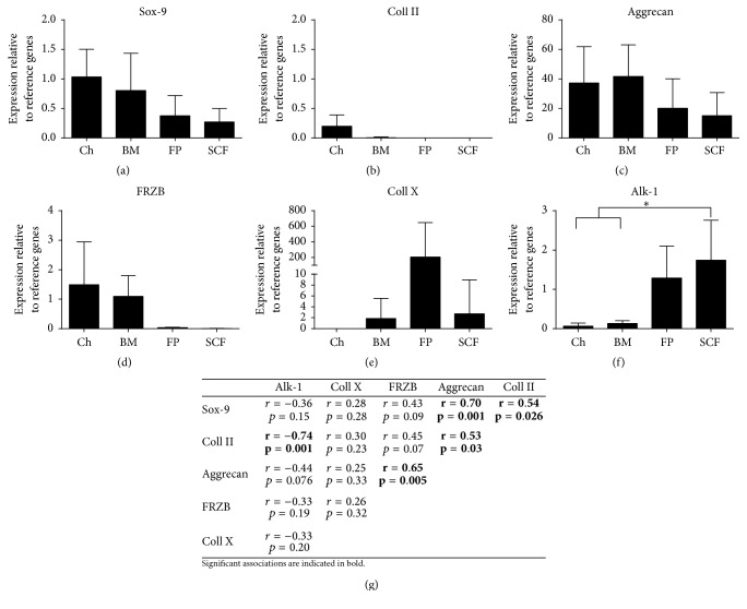
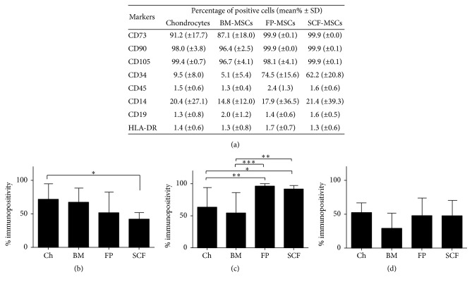
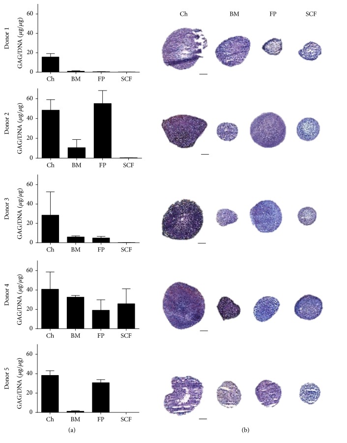
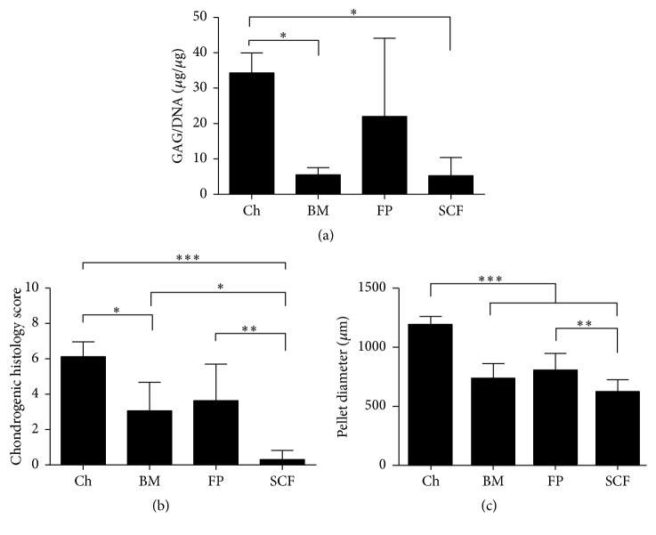
Autologous chondrocyte implantation (ACI) is a cell-based therapy that has been used clinically for over 20 years to treat cartilage injuries more efficiently in order to negate or delay the need for joint replacement surgery. In this time, very little has changed in the ACI procedure, but now many centres are considering or using alternative cell sources for cartilage repair, in particular mesenchymal stem cells (MSCs). In this study, we have tested the chondrogenic potential of donor-matched MSCs derived from bone marrow (BM), infrapatellar fat pad (FP), and subcutaneous fat (SCF), compared to chondrocytes. We have confirmed that there is a chondrogenic potency hierarchy ranging across these cell types, with the most potent being chondrocytes, followed by FP-MSCs, BM-MSCs, and lastly SCF-MSCs. We have also examined gene expression and surface marker profiles in a predictive model to identify cells with enhanced chondrogenic potential. In doing so, we have shown that Sox-9, Alk-1, and Coll X expressions, as well as immunopositivity for CD49c and CD39, have predictive value for all of the cell types tested in indicating chondrogenic potency. The findings from this study have significant clinical implications for the refinement and development of novel cell-based cartilage repair strategies.


Figures

Figure 1
Figure 1
The expression of chondrogenic and hypertrophic genes in monolayer cell populations prior to chondrogenesis. ((a)–(f)) Chondrocytes (Ch), bone marrow MSC (BM), fat pad MSC (FP), and subcutaneous fat MSC (SCF). Data shown are the means ± the standard deviation of triplicate runs and 5 donors for each cell population. One-way ANOVA and post hoc Bonferroni tests were used to test for significant differences in gene expression levels between cell types. (g) Pearson's correlation analysis matrix comparing genes which may be predictive of chondrogenic potential; significant correlations are in bold. Gene expression is expressed relative to the reference genes PPIA and TBP.
Figure 2
Figure 2
Immunoprofiles for MSC markers and putative chondrogenic potency markers of culture expanded cells prior to chondrogenesis. (a) ISCT MSC immunoprofiles. Immunoprofiles for the putative chondrogenic markers CD49c (b), CD166 (c), and CD39 (d). Flow cytometry was used to detect the percentage of positive cells for each marker on monolayer cell populations of chondrocytes (Ch), bone marrow MSC (BM), fat pad MSC (FP), and subcutaneous fat MSC (SCF) prior to chondrogenesis. Data shown are the means ± the standard deviation of 5 donors for each cell population. One-way ANOVA and post hoc Bonferroni tests were used to test for significant differences in the positivity of cell surface markers between cell types.
Figure 3
Figure 3
Chondrogenic assessments of pellet cultures between donors. (a) Production of GAG/DNA in pellet cultures from chondrocytes (Ch), bone marrow MSC (BM), fat pad MSC (FP), and subcutaneous fat MSC (SCF). GAGs were measured after chondrogenic differentiation using the DMMB assay and normalised to the DNA content of pellets; each donor is represented in individual graphs. Data shown are the means ± the standard deviation of triplicate pellets. (b) Chondrogenic pellets from Ch, BM, FP, and SCF showing representative toluidine blue staining for each donor. Scale bars represent 200 μm.
Figure 4
Figure 4
Chondrogenic assessments of pellet cultures across cell types. (a) Production of GAG/DNA in pellet cultures from chondrocytes (Ch), bone marrow MSC (BM), fat pad MSC (FP), and subcutaneous fat MSC (SCF) for all donors combined. (b) Chondrogenic histology scores for Ch, BM, FP, and SCF for all donors combined. (c) Mean chondrogenic pellet diameter (μm) for Ch, BM, FP, and SCF for all donors combined. Data shown are the means ± the standard deviation of triplicate pellets and 5 donors for each cell population. One-way ANOVA and post hoc Bonferroni tests were used to test for significant differences between cell types.

References

    1. Brittberg M., Lindahl A., Nilsson A., Ohlsson C., Isaksson O., Peterson L. Treatment of deep cartilage defects in the knee with autologous chondrocyte transplantation. The New England Journal of Medicine. 1994;331(14):889–895. doi: 10.1056/nejm199410063311401. - DOI - PubMed
    1. Bhosale A. M., Kuiper J. H., Johnson W. E. B., Harrison P. E., Richardson J. B. Midterm to long-term longitudinal outcome of autologous chondrocyte implantation in the knee joint: a multilevel analysis. The American Journal of Sports Medicine. 2009;37(supplement 1):131S–138S. - PubMed
    1. Richardson J. B., Caterson B., Evans E. H., Ashton B. A., Roberts S. Repair of human articular cartilage after implantation of autologous chondrocytes. Journal of Bone and Joint Surgery—Series B. 1999;81(6):1064–1068. doi: 10.1302/0301-620x.81b6.9343. - DOI - PubMed
    1. Saris D. B. F., Vanlauwe J., Victor J., et al. Treatment of symptomatic cartilage defects of the knee: characterized chondrocyte implantation results in better clinical outcome at 36 months in a randomized trial compared to microfracture. The American Journal of Sports Medicine. 2009;37(supplement 1):10S–19S. - PubMed
    1. McCarthy H. S., Richardson J. B., Parker J. C., Roberts S. Evaluating joint morbidity after chondral harvest for autologous chondrocyte implantation (ACI): a study of ACI-treated ankles and hips with a knee chondral harvest. Cartilage. 2016;7(1):7–15. doi: 10.1177/1947603515607963. - DOI - PMC - PubMed

LinkOut - more resources