

doi: 10.3389/fvets.2016.00091.
eCollection 2016.
Detection of Astrovirus in Historical Cases of European Sporadic Bovine Encephalitis, Switzerland 1958-1976
Affiliations
- PMID: 27781208
- PMCID: PMC5058262
- DOI: 10.3389/fvets.2016.00091
Item in Clipboard
Detection of Astrovirus in Historical Cases of European Sporadic Bovine Encephalitis, Switzerland 1958-1976
Front Vet Sci.
.
Abstract
European sporadic bovine encephalitis is a frequent diagnosis in neurologically diseased cattle, but its etiology remained unresolved. Using in situ hybridization, we have detected a recently discovered neurotropic bovine astrovirus in historical tissues in a high proportion of brain samples of affected cattle. Our results suggest that astroviruses were already involved in the pathogenesis of the disease several decades ago, but have gone undetected.
Keywords: archive; astrovirus; cattle; encephalitis; historical; neurological disease.
Figures
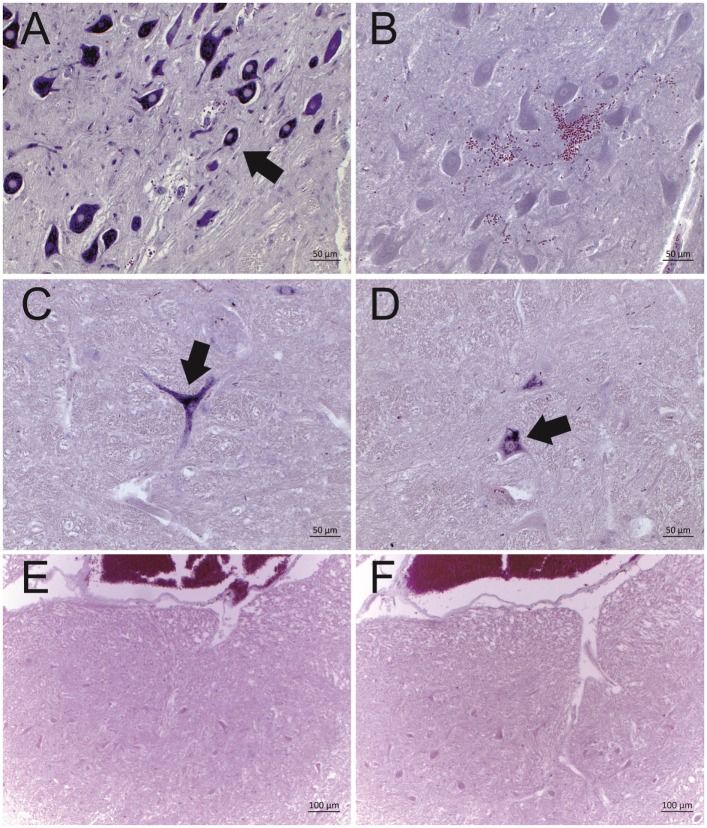
Representative results of the in situ hybridization (ISH) for BoAstV-CH13/NeuroS1 RNA in cattle brain tissues. Two DIG labeled RNA probes [probe A: micrographs (A,C,E); probe B, micrographs (B,D,F)] were applied. Arrows indicate individual ISH-positive neurons. (A,B) Historical case of European sporadic bovine encephalitis (case 3628), which was diagnosed in 1959. Note that only probe A was reactive, but not probe B. (C,D) BoAstV-CH13/NeuroS1 positive control case, dating from 1995. Both probes show a clear labeling of neurons. (E,F) Negative control tissues with the absence of labeling by both probes.
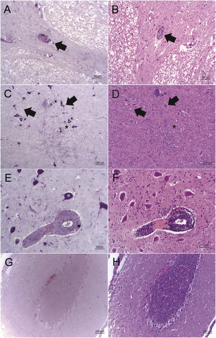
Bovine astrovirus RNA and its correlation with histopathological lesions in brain tissues of historical cases of European sporadic bovine encephalitis. Astrovirus RNA was detected by in situ hybridization with probe A [ISH, purple labeling; (A,C,E,G)] and histopathological lesions were assessed after hematoxylin and eosin staining [HE; (B,D,F,H)]. (A,B) Case 11930, brainstem: good correlation of ISH labeling and histopathological lesions in perivascular cuffs (arrow). (C,D) Case 3628, brainstem: strong ISH labeling in neurons in vicinity of lesions (arrows), and weak ISH labeling of glia cells in a glial node (asterisk). (E,F) Case 3628, brainstem: strongly ISH labeled neurons and weakly labeled mononuclear cells in a perivascular cuff. (G,H) Case 11646, cerebellar cortex: lack of correlation between lesions and ISH labeling. While there is a moderate gliosis, cells remain ISH negative. Magnifications of microphotographs are indicated by scale bars.
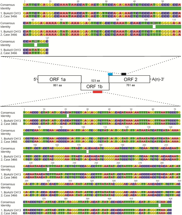
Results of RT-PCR and sequencing of astrovirus RNA extracted from case 3466. A scheme of the BoAstV CH13 genome is presented. The viral genome is organized in three open-reading frames (ORF). The red bar indicates the target sequence for RT-PCR using primers MA2/MA4 within ORF 1b, which encodes for the RNA-dependent RNA polymerase. The blue and black bars show target sequences of in situ hybridization probes A and B, respectively, within ORF2 that encodes for structural capsid proteins. The nested RT-PCR protocol with primers bAV3/bAV4 (first round) and bAV1/bAV2 (second round) was designed to yield amplicons in the target region of ISH probe B (black bar). Sequence comparisons of the MA2/MA4 and of the bAV1/bAV2 amplicons of case 3466 with the BoAstV CH13 reference sequence (GenBank accession number: NC_024498) are shown. Alignments were generated with the Geneious R9 software, version 9.0.4 (Biomatters).
Similar articles
-
Frequency and Pathological Phenotype of Bovine Astrovirus CH13/NeuroS1 Infection in Neurologically-Diseased Cattle: Towards Assessment of Causality.Viruses. 2017 Jan 18;9(1):12. doi: 10.3390/v9010012. Viruses. 2017. PMID: 28106800 Free PMC article.
-
Full-genome based molecular characterization of encephalitis-associated bovine astroviruses.Infect Genet Evol. 2016 Oct;44:162-168. doi: 10.1016/j.meegid.2016.06.052. Epub 2016 Jul 1. Infect Genet Evol. 2016. PMID: 27378415
-
[Neurotropic bovine astrovirus-associated encephalitis: An underdiagnosed disease in South America?].Rev Argent Microbiol. 2022 Apr-Jun;54(2):100-105. doi: 10.1016/j.ram.2021.01.006. Epub 2021 Jun 18. Rev Argent Microbiol. 2022. PMID: 34148730 Spanish.
-
Epidemiology of Classic and Novel Human Astrovirus: Gastroenteritis and Beyond.Viruses. 2017 Feb 18;9(2):33. doi: 10.3390/v9020033. Viruses. 2017. PMID: 28218712 Free PMC article. Review.
-
The changing epidemiology of astrovirus-associated gastroenteritis: a review.Arch Virol Suppl. 1996;12:287-300. doi: 10.1007/978-3-7091-6553-9_31. Arch Virol Suppl. 1996. PMID: 9015126 Review.
Cited by
-
Accurate and precise real-time RT-PCR assays for the identification of astrovirus associated encephalitis in cattle.Sci Rep. 2018 Jun 15;8(1):9215. doi: 10.1038/s41598-018-27533-8. Sci Rep. 2018. PMID: 29907784 Free PMC article.
-
Differential In Vitro Infection of Neural Cells by Astroviruses.mBio. 2019 Jul 9;10(4):e01455-19. doi: 10.1128/mBio.01455-19. mBio. 2019. PMID: 31289185 Free PMC article.
-
The First Case of Bovine Astrovirus-Associated Encephalitis in the Southern Hemisphere (Uruguay), Uncovers Evidence of Viral Introduction to the Americas From Europe.Front Microbiol. 2019 Jun 4;10:1240. doi: 10.3389/fmicb.2019.01240. eCollection 2019. Front Microbiol. 2019. PMID: 31231334 Free PMC article.
-
Neurologic Clinical Signs in Cattle With Astrovirus-Associated Encephalitis.J Vet Intern Med. 2017 Jul;31(4):1209-1214. doi: 10.1111/jvim.14728. Epub 2017 May 22. J Vet Intern Med. 2017. PMID: 28544318 Free PMC article.
-
The Broad Host Range and Genetic Diversity of Mammalian and Avian Astroviruses.Viruses. 2017 May 10;9(5):102. doi: 10.3390/v9050102. Viruses. 2017. PMID: 28489047 Free PMC article. Review.
References
-
- Frauchiger E, Hofmann W. Nervous Diseases of Cattle. Bern: Medizinischer Verlag Hans Huber; (1941).
-
- Fankhauser R. Sporadische meningo-enzephalomyelitis beim Rind. Schweiz Arch Tierheilk (1961) 103:225–35.
-
- Fankhauser R. Cerebelläre Enzephalitis beim Rind. Schweiz Arch Tierheilk (1961) 103(6):11.
-
- Fatzer R, Steck F. [Histological differential diagnosis in cattle suspected of rabies]. Schweiz Arch Tierheilkd (1974) 116(7):347–56. - PubMed
-
- Bestetti G, Fatzer R, Fankhauser R. Ultrastructural investigations concerning a sporadically occurring meningo-encephalomyelitis of cattle in Switzerland. Schweiz Arch Tierheilkd (1976) 118:351–7. - PubMed
LinkOut - more resources
Full Text Sources
Other Literature Sources