

Robust dynamic myocardial perfusion CT deconvolution for accurate residue function estimation via adaptive-weighted tensor total variation regularization: a preclinical study
- PMID: 27782004
- PMCID: PMC5088789
- DOI: 10.1088/0031-9155/61/22/8135
Robust dynamic myocardial perfusion CT deconvolution for accurate residue function estimation via adaptive-weighted tensor total variation regularization: a preclinical study
Abstract
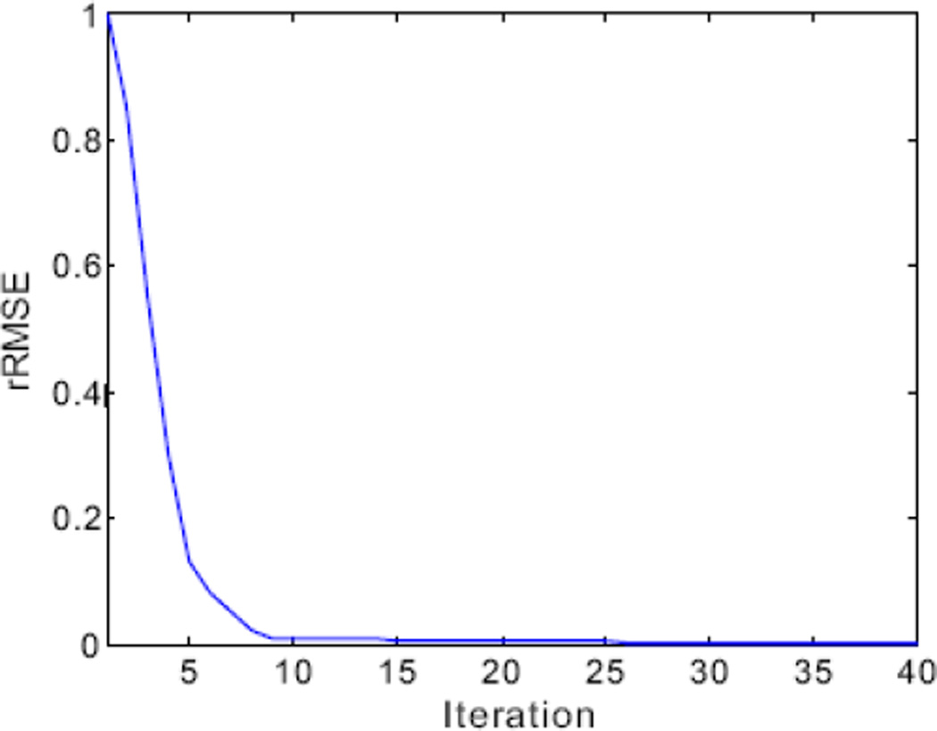
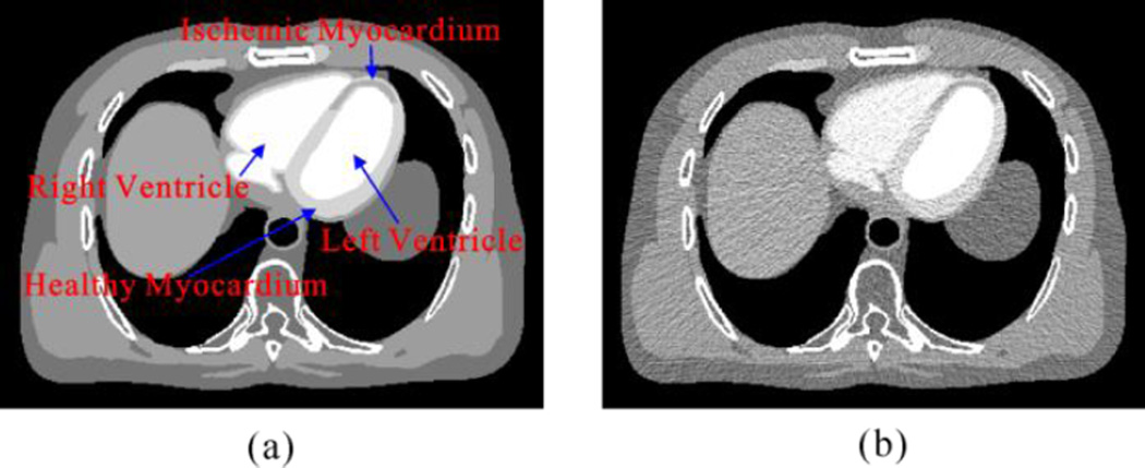
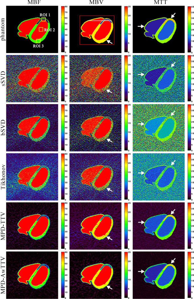
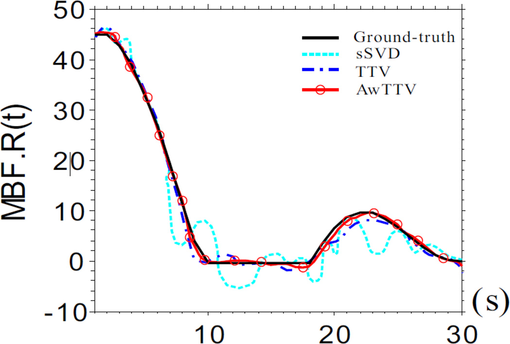
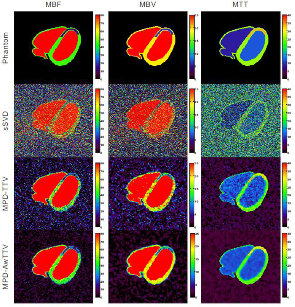
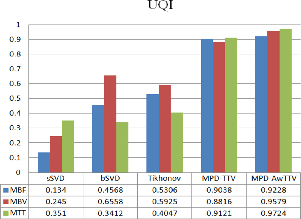
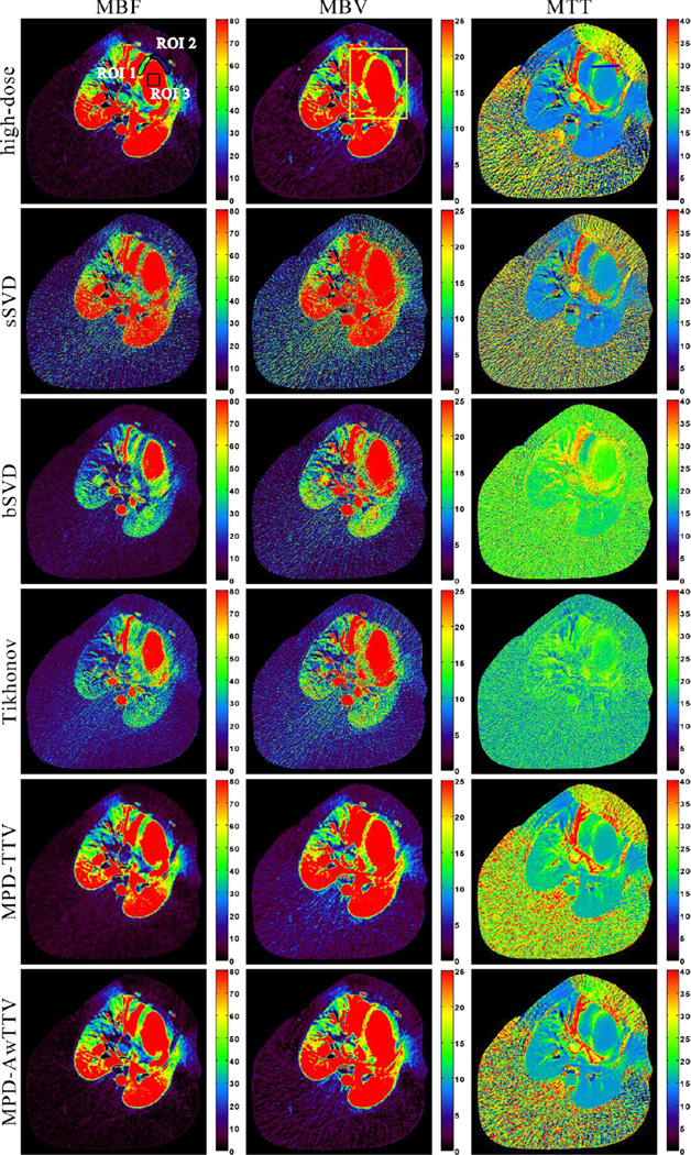
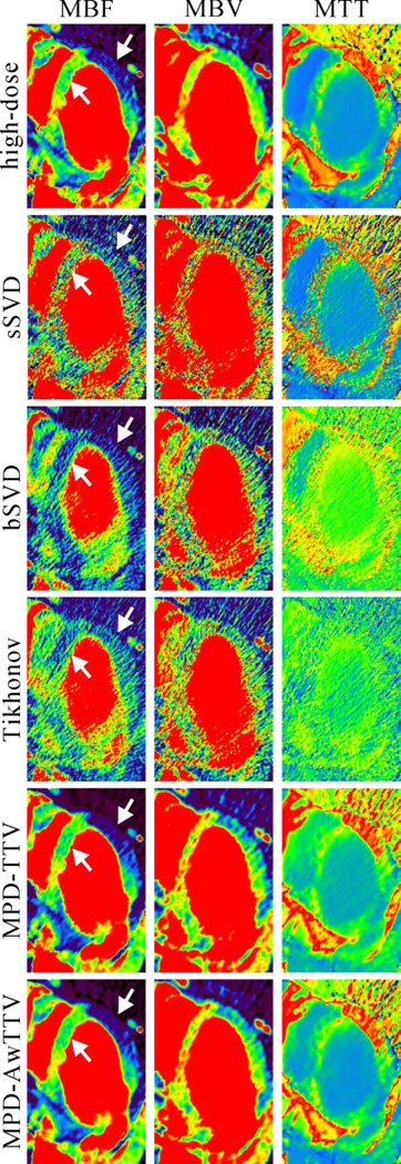
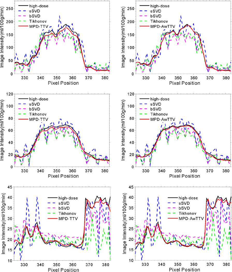
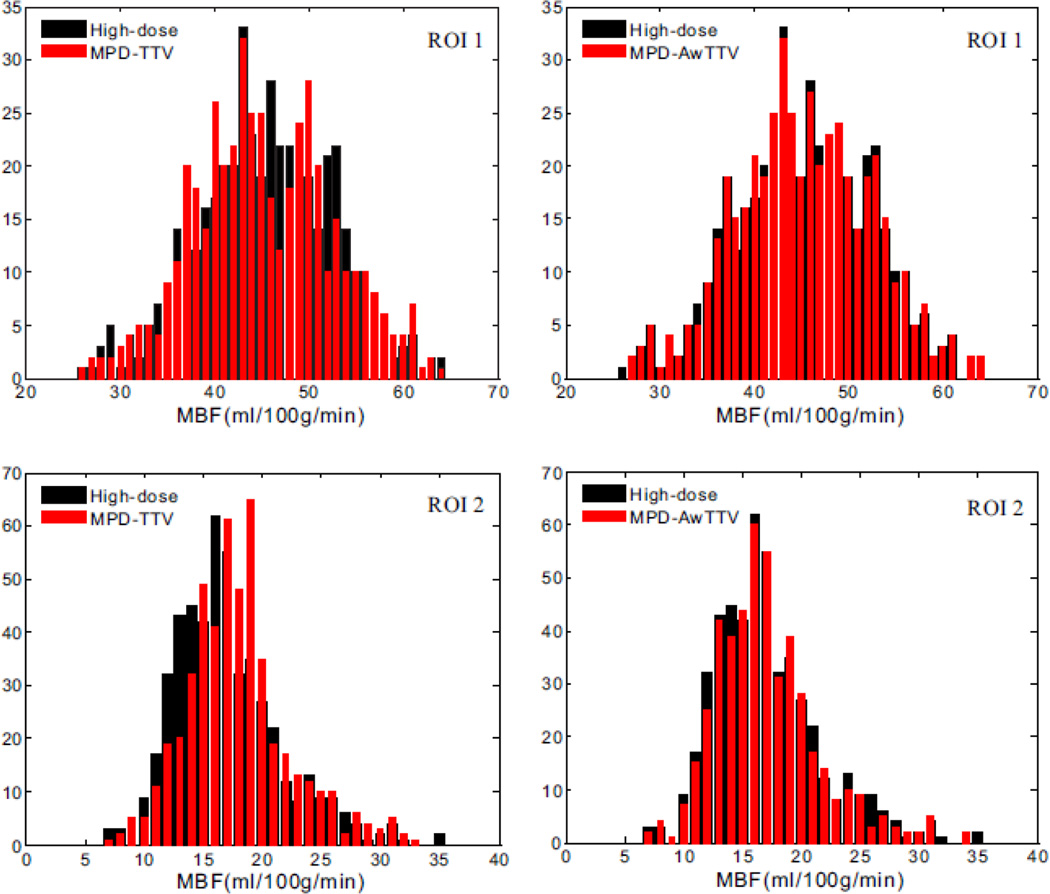
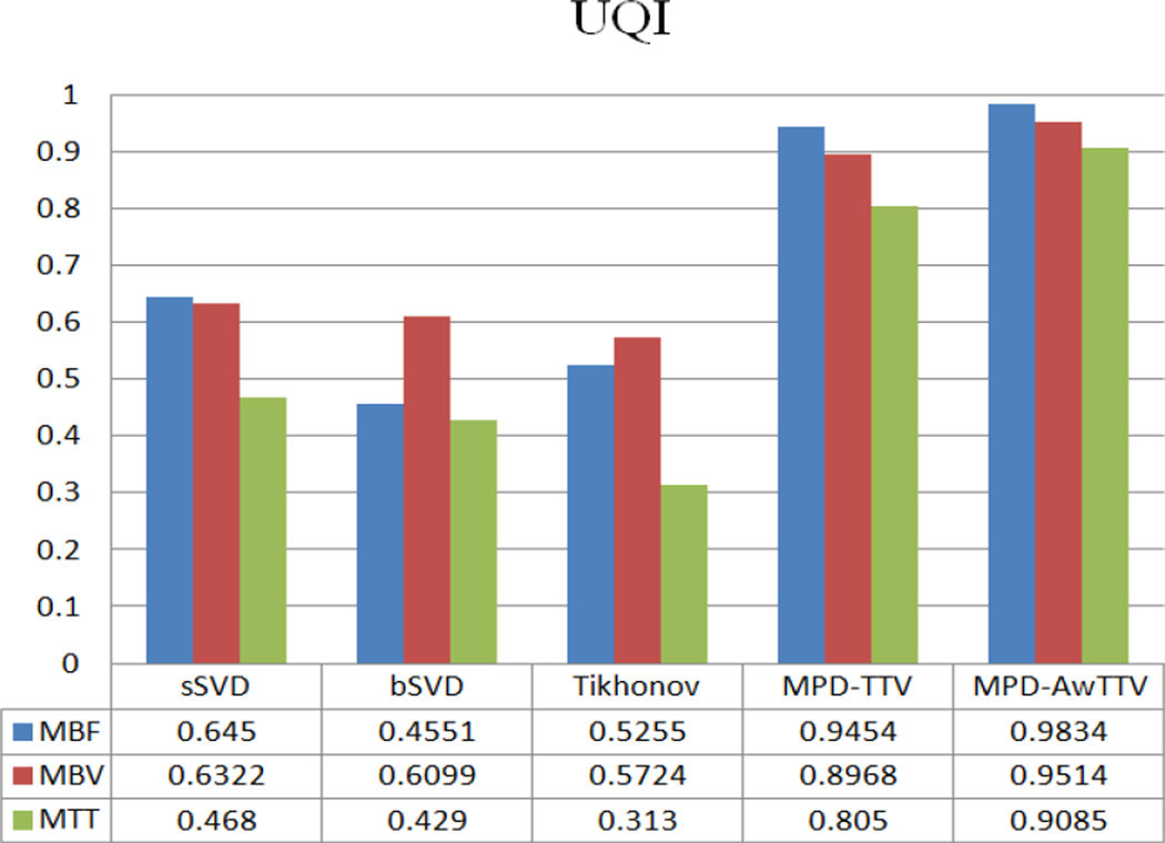
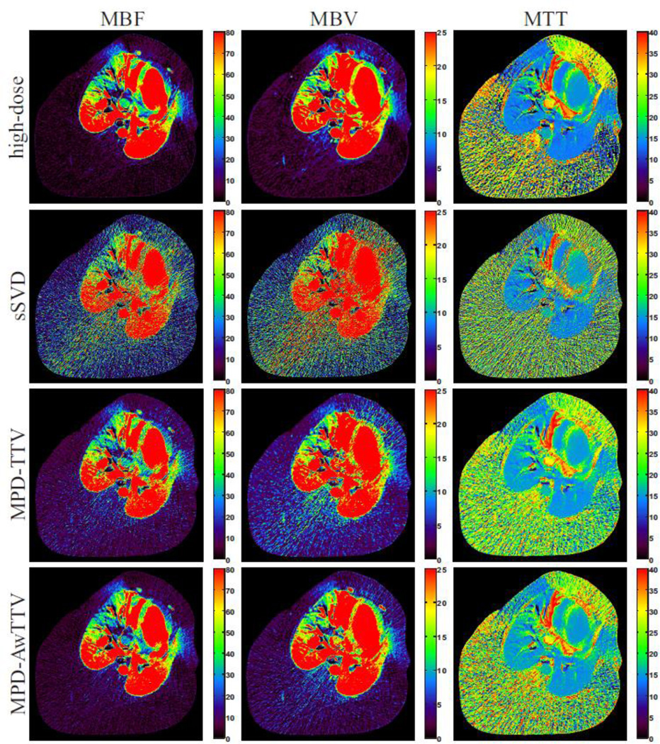
Dynamic myocardial perfusion computed tomography (MPCT) is a promising technique for quick diagnosis and risk stratification of coronary artery disease. However, one major drawback of dynamic MPCT imaging is the heavy radiation dose to patients due to its dynamic image acquisition protocol. In this work, to address this issue, we present a robust dynamic MPCT deconvolution algorithm via adaptive-weighted tensor total variation (AwTTV) regularization for accurate residue function estimation with low-mA s data acquisitions. For simplicity, the presented method is termed 'MPD-AwTTV'. More specifically, the gains of the AwTTV regularization over the original tensor total variation regularization are from the anisotropic edge property of the sequential MPCT images. To minimize the associative objective function we propose an efficient iterative optimization strategy with fast convergence rate in the framework of an iterative shrinkage/thresholding algorithm. We validate and evaluate the presented algorithm using both digital XCAT phantom and preclinical porcine data. The preliminary experimental results have demonstrated that the presented MPD-AwTTV deconvolution algorithm can achieve remarkable gains in noise-induced artifact suppression, edge detail preservation, and accurate flow-scaled residue function and MPHM estimation as compared with the other existing deconvolution algorithms in digital phantom studies, and similar gains can be obtained in the porcine data experiment.
Figures

Similar articles
-
Promote quantitative ischemia imaging via myocardial perfusion CT iterative reconstruction with tensor total generalized variation regularization.Phys Med Biol. 2018 Jun 12;63(12):125009. doi: 10.1088/1361-6560/aac7bd. Phys Med Biol. 2018. PMID: 29794346
-
Cerebral perfusion computed tomography deconvolution via structure tensor total variation regularization.Med Phys. 2016 May;43(5):2091. doi: 10.1118/1.4944866. Med Phys. 2016. PMID: 27147322
-
Low-dose dynamic myocardial perfusion CT image reconstruction using pre-contrast normal-dose CT scan induced structure tensor total variation regularization.Phys Med Biol. 2017 Apr 7;62(7):2612-2635. doi: 10.1088/1361-6560/aa5d40. Epub 2017 Jan 31. Phys Med Biol. 2017. PMID: 28140366
-
Low-dose dynamic myocardial perfusion CT imaging using a motion adaptive sparsity prior.Med Phys. 2017 Sep;44(9):e188-e201. doi: 10.1002/mp.12285. Med Phys. 2017. PMID: 28901610
-
Low-Dose Dynamic Cerebral Perfusion Computed Tomography Reconstruction via Kronecker-Basis-Representation Tensor Sparsity Regularization.IEEE Trans Med Imaging. 2017 Dec;36(12):2546-2556. doi: 10.1109/TMI.2017.2749212. Epub 2017 Sep 4. IEEE Trans Med Imaging. 2017. PMID: 28880164 Free PMC article.
Cited by
-
Statistical CT reconstruction using region-aware texture preserving regularization learning from prior normal-dose CT image.Phys Med Biol. 2018 Nov 20;63(22):225020. doi: 10.1088/1361-6560/aaebc9. Phys Med Biol. 2018. PMID: 30457116 Free PMC article.
-
Contrast-Medium Anisotropy-Aware Tensor Total Variation Model for Robust Cerebral Perfusion CT Reconstruction with Low-Dose Scans.IEEE Trans Comput Imaging. 2020;6:1375-1388. doi: 10.1109/tci.2020.3023598. Epub 2020 Sep 11. IEEE Trans Comput Imaging. 2020. PMID: 33313342 Free PMC article.
-
Robust Low-Dose CT Sinogram Preprocessing via Exploiting Noise-Generating Mechanism.IEEE Trans Med Imaging. 2017 Dec;36(12):2487-2498. doi: 10.1109/TMI.2017.2767290. IEEE Trans Med Imaging. 2017. PMID: 29192885 Free PMC article.
-
[Redundancy information-induced image reconstruction for low-dose myocardial perfusion computed tomography].Nan Fang Yi Ke Da Xue Xue Bao. 2018 Jan 30;38(1):27-33. doi: 10.3969/j.issn.1673-4254.2018.01.05. Nan Fang Yi Ke Da Xue Xue Bao. 2018. PMID: 33177030 Free PMC article. Chinese.
-
Application of the 4-D XCAT Phantoms in Biomedical Imaging and Beyond.IEEE Trans Med Imaging. 2018 Mar;37(3):680-692. doi: 10.1109/TMI.2017.2738448. Epub 2017 Aug 10. IEEE Trans Med Imaging. 2018. PMID: 28809677 Free PMC article. Review.
References
-
- Achenbach S, Goroll T, Seltmann M, Pflederer T, Anders K, Ropers D, Daniel WG, Uder M, Lell M, Marwan M. Detection of coronary artery stenoses by low-dose, prospectively ECG-triggered, high-pitch spiral coronary CT angiography. JACC: Cardiovasc. Imaging. 2011;4:328–337. - PubMed
-
- Beck A, Teboulle M. A fast iterative shrinkage-thresholding algorithm for linear inverse problems. SIAM J. Imaging Sci. 2009;2:183–202.
-
- Becker A, Becker C. CT imaging of myocardial perfusion: possibilities and perspectives. J. Nucl. Cardiol. 2013;20:289–296. - PubMed
-
- Bian Z, Gong C, Zeng D, Niu S, Zhang H, Zhang Z, Ma J. Fully Three-Dimensional Image Reconstruction in Radiology and Nuclear Medicine, Fully 3D Image Reconstruction in Radiology and Nuclear Medicine. Newport, Rhode Island, USA: 2015. [31 May-5 June 2015]. Motion Adaptive Sparsity Prior for Low-dose Dynamic CT Myocardial Perfusion Imaging.
MeSH terms
Grants and funding
LinkOut - more resources
Full Text Sources
Other Literature Sources
Medical