. 2016 Oct 26:7:13207.
doi: 10.1038/ncomms13207.

Wnt and Neuregulin1/ErbB signalling extends 3D culture of hormone responsive mammary organoids

Affiliations

Wnt and Neuregulin1/ErbB signalling extends 3D culture of hormone responsive mammary organoids

Thierry Jardé et al. Nat Commun. .

Abstract

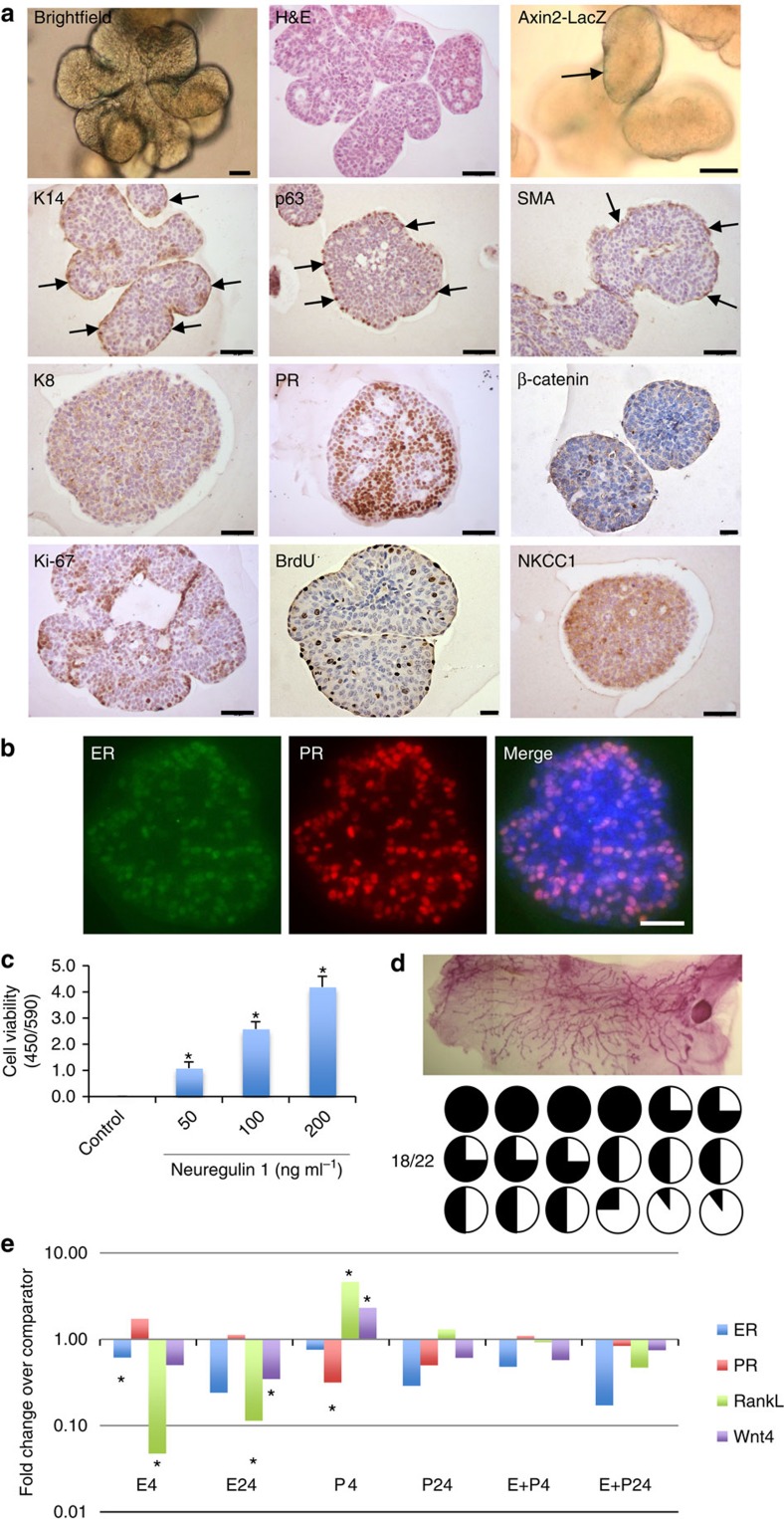
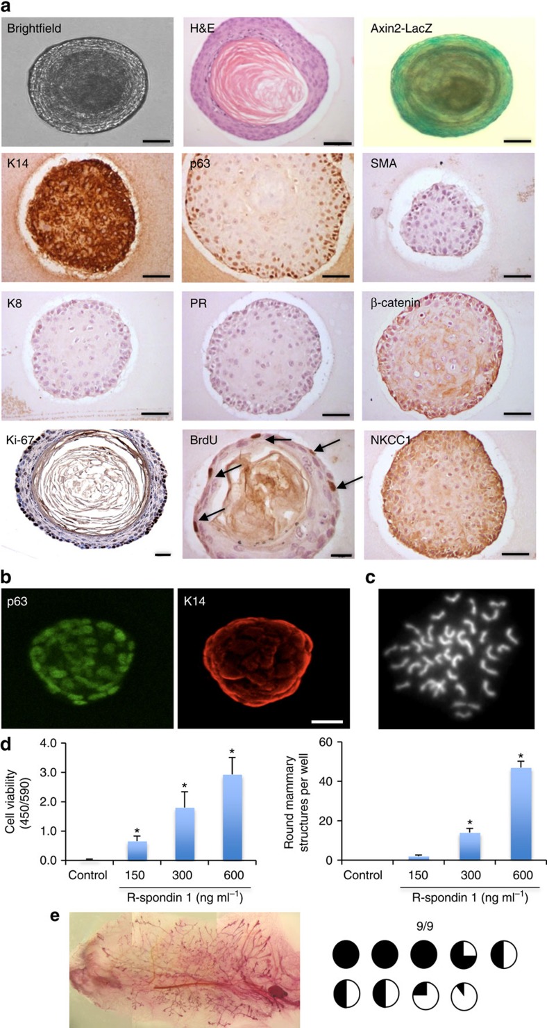
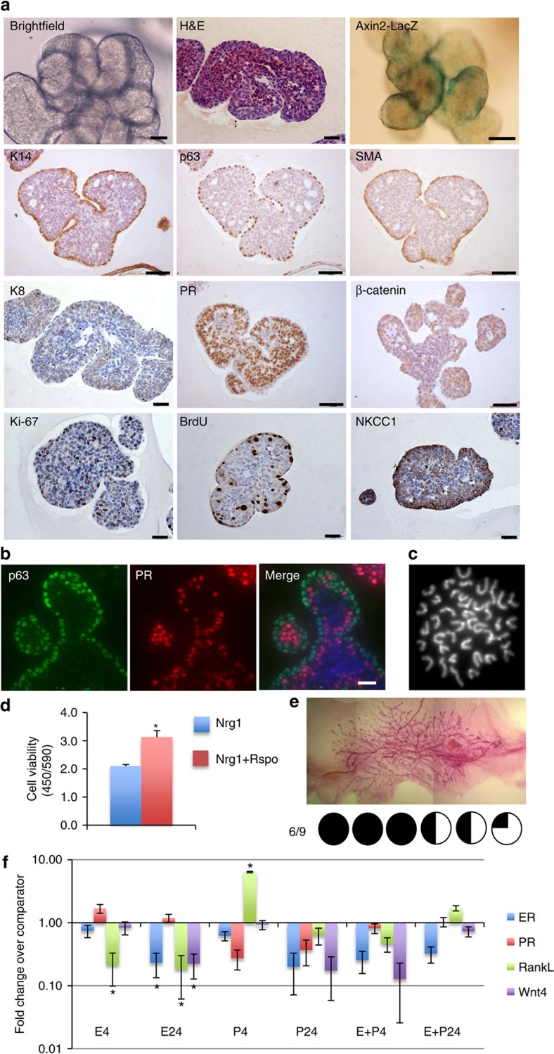
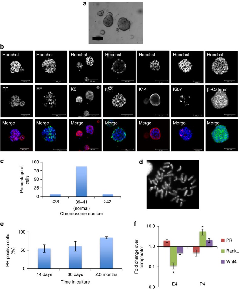
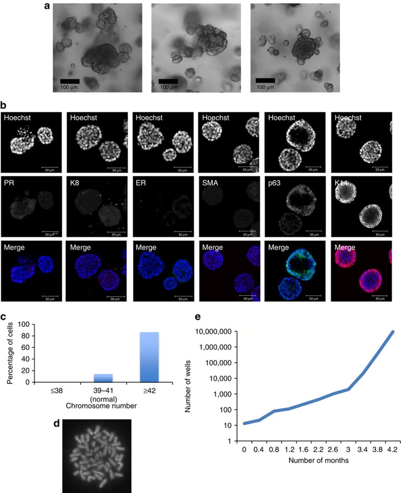
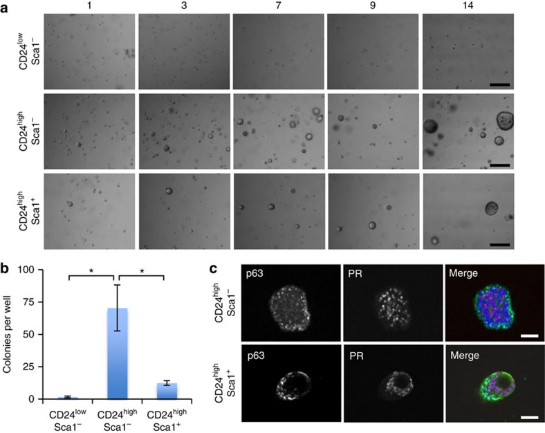
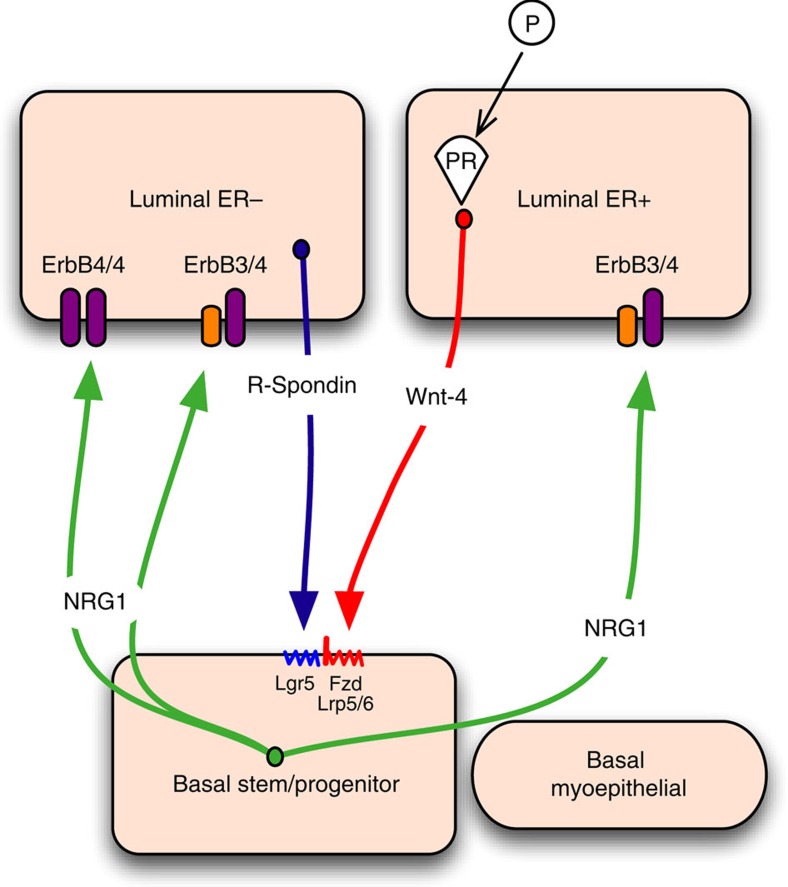
The development of in vitro culture systems quantitatively and qualitatively recapitulating normal breast biology is key to the understanding of mammary gland biology. Current three-dimensional mammary culture systems have not demonstrated concurrent proliferation and functional differentiation ex vivo in any system for longer than 2 weeks. Here, we identify conditions including Neuregulin1 and R-spondin 1, allowing maintenance and expansion of mammary organoids for 2.5 months in culture. The organoids comprise distinct basal and luminal compartments complete with functional steroid receptors and stem/progenitor cells able to reconstitute a complete mammary gland in vivo. Alternative conditions are also described that promote enrichment of basal cells organized into multiple layers surrounding a keratinous core, reminiscent of structures observed in MMTV-Wnt1 tumours. These conditions comprise a unique tool that should further understanding of normal mammary gland development, the molecular mechanism of hormone action and signalling events whose deregulation leads to breast tumourigenesis.


Figures

Figure 1
Figure 1. Nrg1 mediates the extended growth of mammary organoids that retain a bilayered organization and response to steroid hormones.
(a) Mammary epithelial cells were embedded in growth-factor reduced matrigel and cultured in basal culture medium supplemented with Noggin (100 ng ml−1) and Nrg1 (100 ng ml−1; n=5). After 30 days in culture, organoids were fixed, embedded in paraffin and sectioned. Sections were stained for hematoxylin and eosin (H&E), basal markers: K14, p63, SMA; luminal markers: K8, PR; cell-proliferation markers: Ki-67 and BrdU; and cell organization markers: β-catenin and NKCC1. Scale bars, 50 μm, except for bright field, β-catenin and BrdU, 25 μm. Mammary organoids established from Axin2-LacZ (Wnt reporter) mammary glands were fixed and stained for β-galactosidase. (b) Detection of ER (green) and PR (red) in mammary organoid section (n=5). Counterstain, DAPI (blue). Scale bar, 50 μm. (c) Mammary cells were exposed to increasing concentrations of Nrg1. After 30 days in culture, the number of viable cells (Wst assay) was measured (n=3, means±s.e.m.). (d) Representative picture of mammary gland successfully filled with a ductal tree. Fifty mammary structures cultured for 30 days were transplanted into mammary fat pads of 3-week-old Rag-1 or FvB mice from which the endogenous epithelium was surgically removed. In all, 18 out of 22 mammary fat pads were re-populated with various degrees (from 5 to 100%). (e) Expression of ER, PR, RankL and Wnt4 following treatment with progesterone (P, 40 ng ml−1), oestrogen (E, 4 ng ml−1) or both hormones. Mammary organoids cultured for 30 days were treated for 4 and 24 h. Gene expression was evaluated by quantitative RT-PCR. Data are expressed as fold change over untreated controls (n=3, means±s.e.m.). *P<0.05, paired Student's t-test.
Figure 2
Figure 2. Nrg1 and ErbB are expressed in distinct mammary cell populations.
Mammary epithelial cell populations were FACS sorted using a panel of antibodies: CD24low Sca-1 CD49fhigh (stem cell-enriched fraction), CD24low Sca-1 CD49flow (myoepithelial cell population), CD24high Sca-1 (luminal ER-negative cells) and CD24high Sca-1+ (luminal ER-positive cells). Expression of Neuregulin 1, Erbb3 and Erbb4 was evaluated by quantitative RT-PCR (n=3 independent experiments). Fold expression±95% confidence over the comparator population (CD24highSca1). ND, no expression of Erbb4 detected. *P<0.05; #0.05<P<0.1 (versus CD24high Sca1), unpaired Student's t-test.
Figure 3
Figure 3. R-spondin 1 promotes the growth of mammary organoids that are enriched in basal cells.
(a) Mammary epithelial cells (n=5) were embedded in growth-factor reduced matrigel. After polymerization of matrigel, basal culture medium supplemented with R-spondin 1 (600 ng ml−1), Noggin (100 ng ml−1) and EGF (50 ng ml−1) was overlaid. After 18 days in culture, organoids were fixed, embedded in paraffin and sectioned. Sections were stained for hematoxylin and eosin (H&E), basal markers: K14, p63, SMA; luminal markers: K8, progesterone receptor (PR); cell-proliferation markers: Ki-67 and BrdU; and cell organization markers: β-catenin and NKCC1. Scale bars, 50 μm, 30 μm (BrdU) and 25 μm (Ki-67). Mammary organoids established from Axin2-LacZ (Wnt reporter) mammary glands were fixed and stained for β-galactosidase. (b) Confocal images of organoids stained for p63 (green) and K14 (red; n=5). Scale bar, 50 μm. (c) Representative picture of metaphase chromosome spread that shows a normal number of chromosomes. (d) Mammary cells were exposed to increasing concentrations of R-spondin 1. After 18 days in culture, cell viability (Wst assay) and the number of structures were evaluated (n=3, means±s.e.m.). (e) Representative picture of mammary gland successfully filled with a ductal tree. Mammary structures cultured for 30 days were transplanted into mammary fat pads of 3-week-old FvB mice from which the endogenous epithelium was surgically removed. In all, 9 out of 9 mammary fat pads were re-populated with various degrees (from 5 to 100%). *P<0.05, paired Student's t-test.
Figure 4
Figure 4. Nrg1 and R-spondin 1 enhance the formation of a basal cell layer while maintaining the luminal compartment.
(a) Mammary epithelial cells were embedded in growth-factor reduced matrigel. After polymerization of matrigel, basal culture medium supplemented with Noggin (100 ng ml−1), R-spondin 1 (100 ng ml−1) and Nrg1 (100 ng ml−1) was overlaid. After 30 days in culture, organoids were fixed, embedded in paraffin and sectioned (n=5). Sections were stained for hematoxylin and eosin (H&E), basal markers: K14, p63, SMA; luminal markers: K8, PR; cell-proliferation markers: Ki-67 and BrdU; and cell-organization markers: β-catenin and NKCC1. Scale bars, 50 μm except for bright field, H&E, K8, Ki-67, BrdU and NKCC1 (25 μm). Mammary organoids established from Axin2-LacZ (Wnt reporter) mammary glands were fixed and stained for β-galactosidase. (b) Detection of p63 (green) and PR (red) in mammary organoid section. Counterstain, DAPI (blue; n=5). Scale bar, 50 μm. (c) Representative picture of metaphase chromosome spread that shows a normal number of chromosomes. (d) Mammary cells were treated with Nrg1 (100 ng ml−1) or Nrg1+R-spondin 1 (100 ng ml−1 each) for 30 days. The number of viable cells (Wst assay) was evaluated (n=3, means±s.e.m.). (e) Representative picture of mammary gland successfully filled with a ductal tree. Mammary structures cultured for 30 days were transplanted into mammary fat pads of 3-week-old FvB mice from which the endogenous epithelium was surgically removed. In all, 6 out of 9 mammary fat pads were re-populated with various degrees (from 15 to 100%). (f) Expression of ER, PR, RankL and Wnt4 following treatment with progesterone (P, 40 ng ml−1), oestrogen (E, 4 ng ml−1) or both hormones. Mammary organoids cultured for 30 days were treated for 4 and 24 h and gene expression evaluated by quantitative RT-PCR. Data are expressed as fold change (versus untreated, n=3, means±s.e.m.). *P<0.05, paired Student's t-test.
Figure 5
Figure 5. Organoids grown for 70 days under Nrg1/Rspo culture conditions retain normal phenotype and functional steroid receptors.
Mammary organoids were cultured with 2.6 ng ml−1 R-spondin 1 (Peprotech), 100 ng ml−1 Nrg1 and 100 ng ml−1 Noggin for 70 days (4 passages). (a) Representative bright field image of the organoids. Scale bar, 100 μm. (b) Organoids were fixed and stained for luminal markers PR, K8 and ER, basal markers K14, p63, and Ki-67 and β-catenin. Counterstain, hoechst (blue). Scale bars, 50 μm. (c) Quantification of chromosome number in mammary organoids cultured for 70 days (n=15). Chromosomal spreads with 40±1 chromosomes were considered normal. (d) Representative picture of metaphase chromosome spread that shows a normal number of chromosomes. (e) Percentage of PR-positive cells per organoid at different time points in culture (n=3). (f) Expression of PR, RankL and Wnt4 following treatment with oestrogen (E, 4 ng ml−1) or progesterone (P, 40 ng ml−1) for 4 h. The gene expression was evaluated by quantitative RT-PCR. Data are expressed as fold change (versus untreated, n=3, means±s.e.m.). *P<0.05, paired Student's t-test.
Figure 6
Figure 6. Long-term culture of mammary organoids with Nrg1 and R-spondin 1 promotes chromosomal abnormalities.
Mammary epithelial cells were freshly isolated, embedded in matrigel, exposed to culture medium containing Nrg1 (100 ng ml−1), Noggin (100 ng ml−1) and R-spondin 1 (2.7 ng ml−1), and cultured for 112 days (passaged 10 times). (a) Representative bright-field images of mammary organoids. Scale bar, 100 μm. (b) Mammary organoids were fixed and stained for the basal markers SMA, p63 and K14, and luminal markers PR, K8 and ER (DAPI, blue). Scale bar, 50 μm. (c) Quantification of chromosome number in mammary organoids (n=29). Chromosomal spreads with 40±1 chromosomes were considered normal. (d) Representative picture of metaphase chromosome spread that shows an aberrant number of chromosomes. (e) Quantification of organoid expansion over time.
Figure 7
Figure 7. Mammary organoid-forming cells are located in the luminal ER-negative cell population and provide all the cell lineages in vitro.
(a) Representative pictures of mammary organoids originating from single CD24high Sca-1 cells (luminal ER-negative fraction), CD24high Sca-1+ cells (luminal ER-positive fraction) or single basal cells (CD24low). Scale bars, 100 μm. (b) Colony-forming efficiency from the three different cell populations (n=3, means±s.e.m.). (c) CD24high Sca1 and CD24high Sca-1+ cells generate mammary organoids that contain distinct luminal and basal compartments (n=3). Scale bars, 50 μm. *P<0.05, paired Student's t-test.
Figure 8
Figure 8. Model of the Wnt/Nrg1 paracrine signalling network between luminal and basal mammary epithelial cells.
Basal cells secrete Nrg1, resulting in Nrg1/ErbB signalling activation in luminal cells, promoting luminal progenitor cell expansion and differentiation. In contrast, progesterone-stimulated Wnt4 secretion by luminal ER+ cells, alongside R-spondin 1 produced by luminal ER cells, activate Wnt signalling in the basal stem/progenitor cell population via the Wnt receptors Fz and Lrp5/6 and the R-spondin 1 receptor Lgr5. Wnt signalling activation in this compartment is likely to promote growth and differentiation of the basal cell layer. Fz, frizzled; Lgr5, leucine-rich repeat-containing G-protein-coupled receptor 5; Lrp5/6, low-density lipoprotein receptor-related protein 5/6.

References

    1. Rios A. C., Fu N. Y., Lindeman G. J. & Visvader J. E. In situ identification of bipotent stem cells in the mammary gland. Nature 506, 322–327 (2014). - PubMed
    1. Van Keymeulen A. et al.. Distinct stem cells contribute to mammary gland development and maintenance. Nature 479, 189–193 (2011). - PubMed
    1. Mroue R. & Bissell M. J. Three-dimensional cultures of mouse mammary epithelial cells. Methods Mol. Biol. 945, 221–250 (2013). - PMC - PubMed
    1. Shamir E. R. & Ewald A. J. Three-dimensional organotypic culture: experimental models of mammalian biology and disease. Nat. Rev. Mol. Cell Biol. 15, 647–664 (2014). - PMC - PubMed
    1. Ewald A. J., Brenot A., Duong M., Chan B. S. & Werb Z. Collective epithelial migration and cell rearrangements drive mammary branching morphogenesis. Dev. Cell 14, 570–581 (2008). - PMC - PubMed

Publication types

MeSH terms